Caso de Estudio
Differential Diagnosis of Glaucoma-like Lesions Using Photopic Negative Response
Dr. Bartłomiej Kocurek y Prof. Adrian Smędowski Universidad Médica de Silesia, Polonia
A 27-year-old male glaucoma suspect was referred to an ophthalmologist to obtain a definitive diagnosis. The patient did not report any visual complaints. His best corrected visual acuity (BCVA) was 20/20 in both eyes. His pressures using the iCare device was 17 mmHg binocularly. During examination of the posterior segment, large optic discs were noted at the level of the fundus, with clear borders and no signs of atrophy.


Desafío
Making a Definitive Diagnosis

DIAGNÓSTICO
Glaucoma

Tipo de evaluación
PhNR (Respuesta Fotópica Negativa)
Why Was the ERG Test Performed?
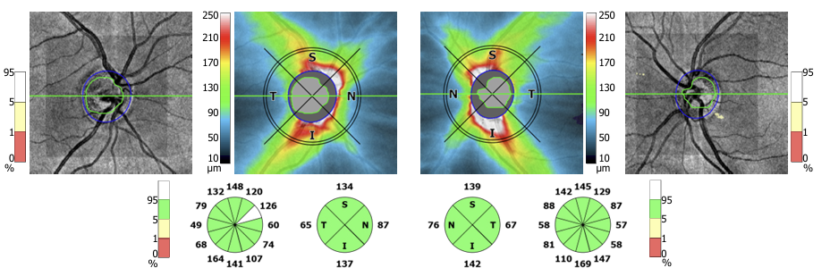
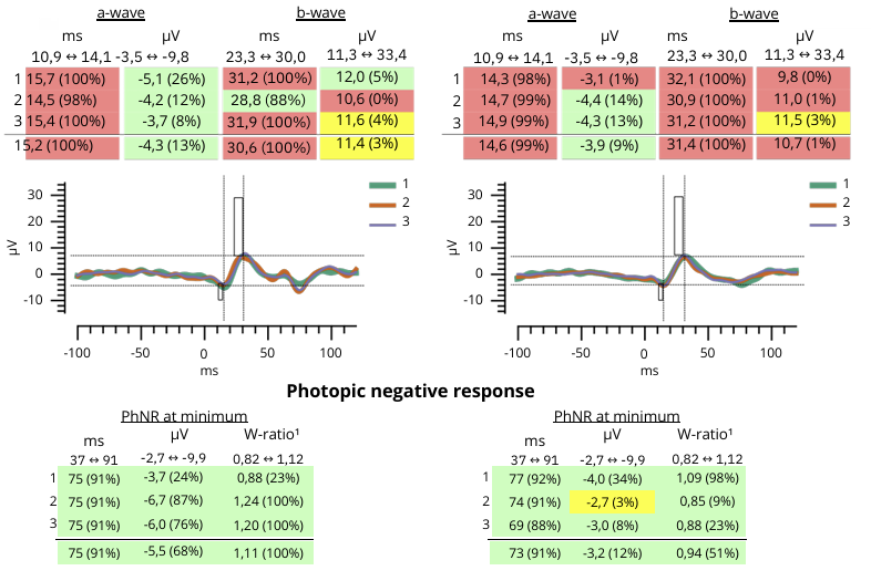
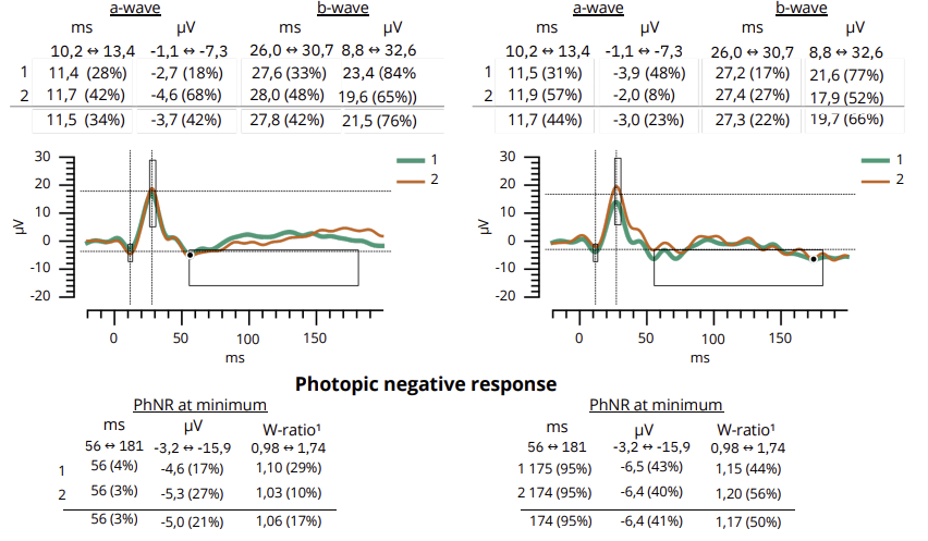
An OCT examination using SD OCT (Revo, Optopol, Poland) was ordered, which showed a normal RNFL profile and thickness (Figure 1). The area of the optic disc OD was 2.68 mm2 and OS was 2.23 mm2, and the c/d ratio OD was 0.54 and OS 0.45 (Figure 2). Humphrey static perimetry was performed using 24-2 SITA strategy, the result of which was within normal limits (Figure 3). To objectively assess the activity of retinal ganglion cells, a photopic negative response (PhNR) test was performed using the RETeval device from LKC Technologies.
Figure 1:
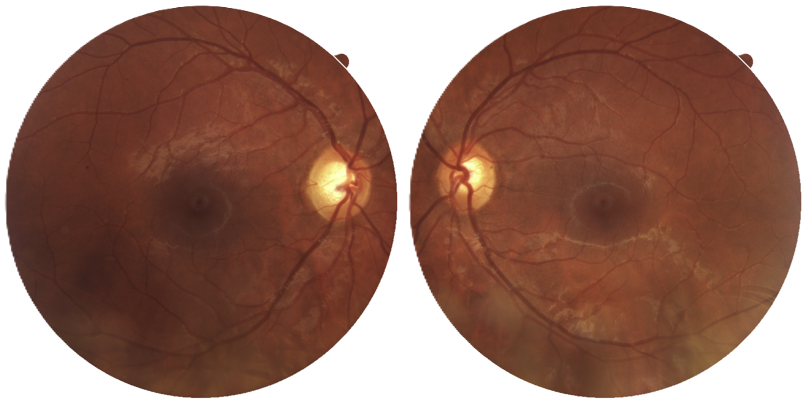
Figure 2:
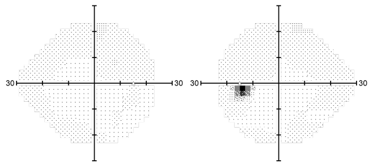
Figure 3:
What Were the ERG Findings?
The PhNR amplitude at minimum for the right eye was -5.5 μV and -3.2 μV for the left eye (Figure 4). These results are within the normal limits.
Figure 4:
How Did ERG Impact the Next Steps?
Normal PhNR results supported the conclusion that the large optic disc represented a variant of normality rather than glaucoma, and observation was initiated. Preventive use of citicoline tablets was recommended. A follow-up visit was performed 2 years later. The BCVA was 20/20. Intraocular pressure was lower at 14 mmHg. OCT (Figure 5) and visual field (Figure 6) results did not reveal any changes, and the PhNR result was -5.0 μV and -6.4 μV for the right and left eyes, respectively (Figure 7).
Figure 5:
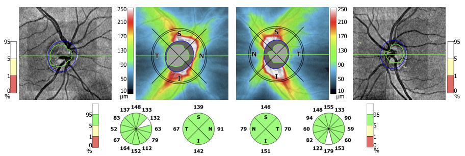
Figure 6:
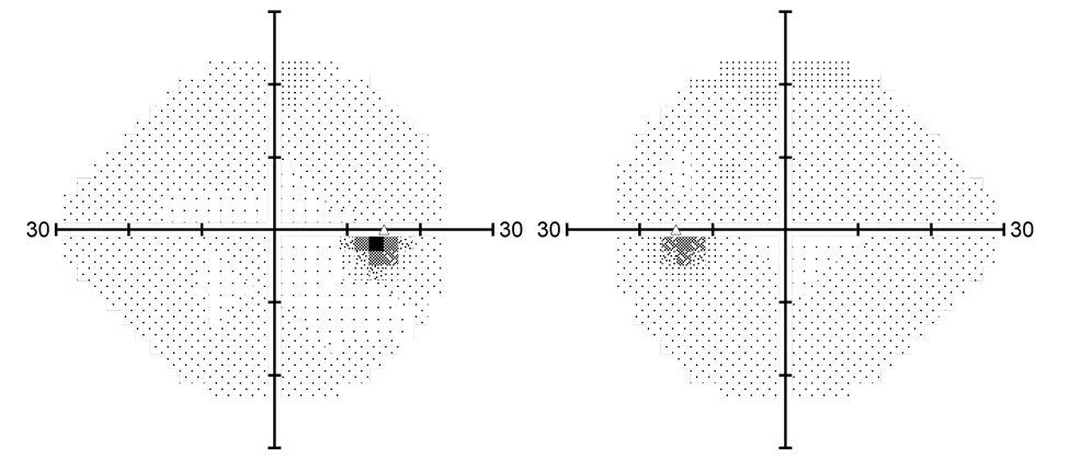
Figure 7:
Conclusión
A large optic disc is an anatomical anomaly but may raise suspicion of glaucoma. Functional assessment of ganglion cells is often a necessary step in differentiating glaucomatous changes from congenital figures.

Dr. Bartłomiej Kocurek u0026 Prof. Adrian Smędowski
Universidad Médica de Silesia.
Dr. Bartłomiej Kocurek was educated at the Medical University of Silesia. After completing his studies, he began work to understand ERG and its utility in managing glaucoma in collaboration with Professor Adrian Smędowski, MD, PhD, FEBO.
Prof. Smędowski is a Professor of Ophthalmology at the Medical University of Silesia in Katowice, Poland, where he serves as Acting Head of the Department of Pediatric Ophthalmology and Deputy Head of the Department of Ophthalmology. He is a Fellow of the European Board of Ophthalmology and a specialist in clinical ophthalmology. Prof. Smędowski earned his medical and doctoral degrees from the Medical University of Silesia and completed postdoctoral research at the University of Eastern Finland. His research focuses on neuroprotective gene therapies for glaucoma and protein interactions in retinal ciliopathies. He leads translational ophthalmology research and is CEO of GlaucoTech and founder of GlaucoMed.