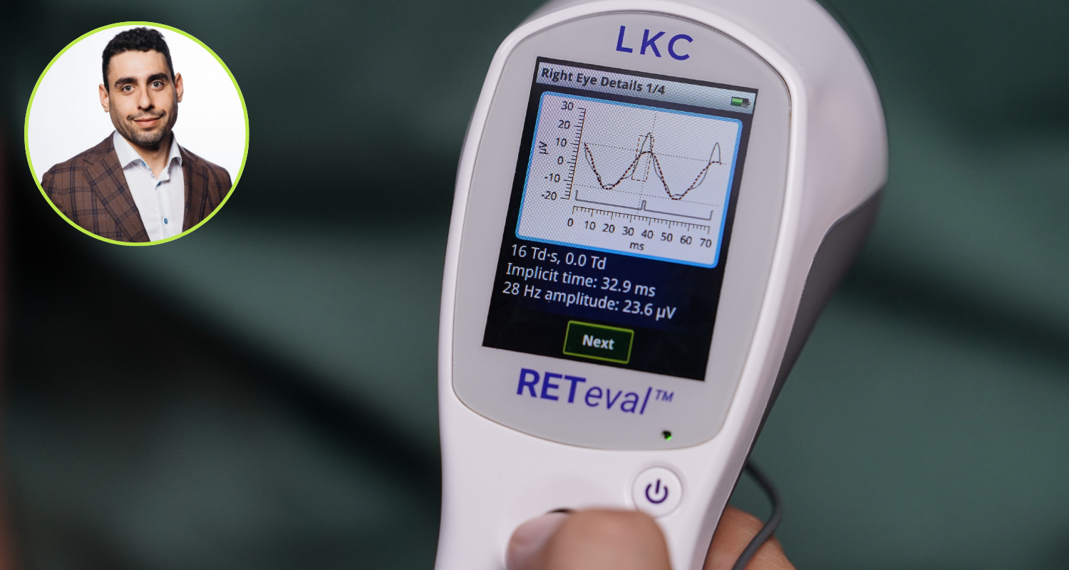
How the RETeval ERG Has Enhanced My Practice
By Jordan Nissinoff, OD

As optometrists, we’re always on the lookout for technologies that can help us provide better care for our patients, especially those with chronic conditions like diabetes, glaucoma, and retinal disease. I’ve found that integrating the RETeval (ERG) into my practice has allowed me to detect and monitor progression more effectively, and I’d like to share my experience with you, along with some key insights into how this technology works.
Why I Chose RETeval ERG for My Practice
Currently, I operate four locations across northern New Jersey, each located inside LensCrafters. I initially started with one office, but as we grew, I was always searching for innovative tools that could elevate our level of care. When I first learned about the RETeval, I saw it as a different tool in the toolbox – one that could potentially help us detect and monitor retinal diseases with more precision.
We’ve now implemented the RETeval in three of our four locations, and I’ve been thoroughly impressed with the difference it has made. Our decision to incorporate it was driven by the desire to provide the most accurate and early detection of pathologies like diabetic retinopathy, glaucoma, and other retinal disorders, which are often challenging to diagnose in their early stages.
What Exactly Is ERG?
For those who may not be as familiar with ERG, it offers a non-invasive, objective measure of retinal function, making it an ideal tool for assessing patients with diabetic retinopathy (DR). Unlike visual acuity and visual field tests, which rely on patient feedback, ERG requires no subjective feedback. Rather, it provides an assessment of retinal health.
In simpler terms, ERG is to the eye what the electrocardiogram (ECG) is to the heart. Just as an ECG is crucial for diagnosing cardiac conditions and monitoring heart function, ERG plays an instrumental role in early detection of retinal dysfunction. This technology allows us to assess the retina’s functionality by capturing electrical signals generated by retinal cells in response to a light stimulus.
When light enters the eye, it’s absorbed by the photoreceptors (rods and cones) in the outer retina. These photoreceptors then convert the light energy into electrical signals, which are transmitted through the inner retinal cells (bipolar and ganglion cells) and eventually travel along the optic nerve to the brain. The ERG captures these electrical responses, allowing us to assess how well different layers of the retina are functioning.
How ERG Complements Structural Imaging
While structural imaging, such as OCT, is crucial for identifying anatomical changes in the retina, ERG offers a functional perspective on retinal health. Functional changes often occur before structural damage is visible. By combining both structural and functional assessments, we gain a more comprehensive understanding of the patient’s retinal health. For example, a patient may show minimal structural changes on OCT but exhibit significant functional deficits on ERG, indicating that the retina is not functioning optimally despite its relatively normal appearance. In such cases, monitoring the patient more closely or referring them to a retinal specialist for further evaluation and potential intervention is warranted. Conversely, a patient with stable retinal function on ERG and minimal structural changes on OCT may not require immediate intervention and can continue with routine monitoring.
The Three Components of Full-Field ERG
El dispositivo ERG/PEV portátil RETeval ERG focuses on three main components in the flash ERG waveform: the a-wave, the b-wave, and the photopic negative response (PhNR). Since each portion of an ERG waveform originates from different layers of the retina, any abnormalities can help pinpoint the site of retinal dysfunction. The two key measurements we analyze are amplitudes (how strong the retinal response is) and implicit times (how fast the retina reacts to the light stimulus). In practice, these measurements help us understand the degree of cellular health. For DR patient management in optometric practice, we focus on the DR Score. This numeric score evaluates the amplitude, timing, and pupillary response to stimuli. A score of 23.5 or above suggests an 11-fold increase in the likelihood that the patient will require retinal intervention (laser or anti-VEGF) within three years. [i]
Implementing ERG in My Practice
We often perform ERG testing on patients with DR, high blood pressure, dark adaptation complaints, or peripheral retinal degeneration. The ERG helps us identify asymmetries between the eyes and provides critical information about cellular activity, making it easier to spot even subtle changes over time.
In terms of practical integration, I’d recommend dedicating time to train your team properly. Like any new technology, there was a learning curve at first, but once my technicians became comfortable with the device, it became a seamless part of our workflow. Now, our technicians handle the testing while the doctors interpret the results, which maintains efficiency without compromising quality care.
The Financial Perspective
It’s also worth noting that incorporating ERG into your practice can be financially rewarding. The test is co-billable with other diagnostic tests such as OCT and fundus photography, and there are 560 ICD-10 codes available for ERG, which further expands its applicability.
Impact on Patient Care
El dispositivo ERG/PEV portátil RETeval has been invaluable in helping us detect changes early on, especially for conditions like diabetic retinopathy and glaucoma. Having this additional data allows us to monitor our patients more closely, adjust treatment plans as needed, and ensure we’re providing the highest level of care possible.
I’ve found that patients appreciate the extra level of detail that ERG testing provides. It helps build trust when we can show them the data and explain what’s happening with their vision at a cellular level. This technology has also improved our ability to track disease progression, which is critical for managing chronic conditions effectively.
Looking Ahead
As optometrists, we have a responsibility to stay on the cutting edge of technology to provide the best care possible for our patients. Incorporating tools like the RETeval into our practices not only enhances our diagnostic capabilities but also reinforces our commitment to delivering comprehensive eye care.

Jordan Nissinoff, OD
Eyewellnis LLC
Dr. Nissinoff is a seasoned optometrist with an expansive background in the optical industry, currently leading a dynamic team at LensCrafters as they develop scalable systems and practices to optimize service delivery. Raised in the optical world, his extensive experience encompasses lab work, eye exams, and navigating insurance intricacies. Dr. Jordan earned his academic foundation in economics from the University of Florida and his doctor of optometry degree from the Pennsylvania College of Optometry, blending clinical expertise with data-driven strategies. Endorsed by industry peers, Dr. Nissinoff’s commitment to excellence sets him apart as a leading contender in the optical world.
