Caso de Estudio
When Standard Tests Aren’t Enough: Using ERG to Support Diagnosis in Case with High Myopia
by Dr. Hussein Almuhtaseb, The View Hospital, Qatar

Desafío
Align Findings

DIAGNÓSTICO
Retinitis Pigmentosa

Testing Protocol
ISCEV 5- Step
Patient History
A 50-year-old female with a history of myopia presented with complaints of progressive vision loss. She previously underwent refractive laser treatment to address visual complaints and manage her myopia. Currently, she reports new-onset vision loss, with visual acuity reduced to 0.9 in the right eye and 0.5 in the left eye.
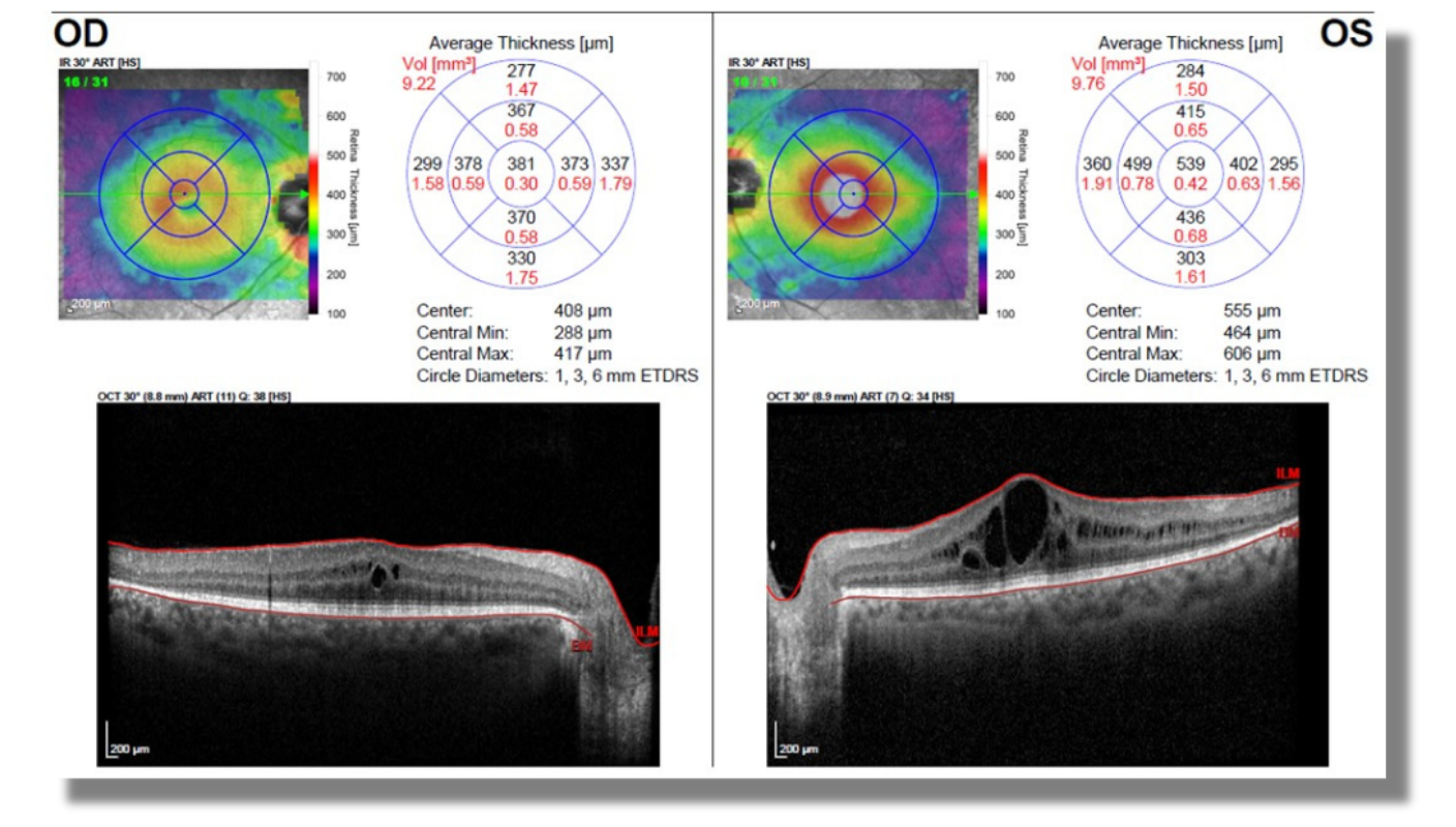
Fundoscopic examination (Figure 1) revealed bone spicule pigmentation affecting the superior posterior pole in both eyes. Visual field testing demonstrated corresponding areas of visual field loss bilaterally. Optical coherence tomography (OCT) confirmed the presence of cystoid macular edema in both eyes (Figure 2), more pronounced in the left. Additional findings included arterial attenuation and optic disc pallor in both eyes.
Figure 1: Fundus Exam
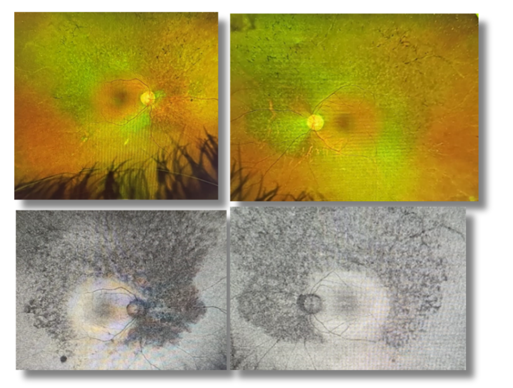
Figure 2: OCT
Why Was an ERG Test Performed?
Due to her progressively worsening vision, which had not responded to previous management strategies, this patient came to us for a second opinion. Based on imaging findings, we suspected inherited retinal degeneration rather than a treatable refractive or structural cause. To further evaluate retinal function and clarify the underlying diagnosis, we recommended a RETeval electroretinogram (ERG).
What Were the ERG Findings?
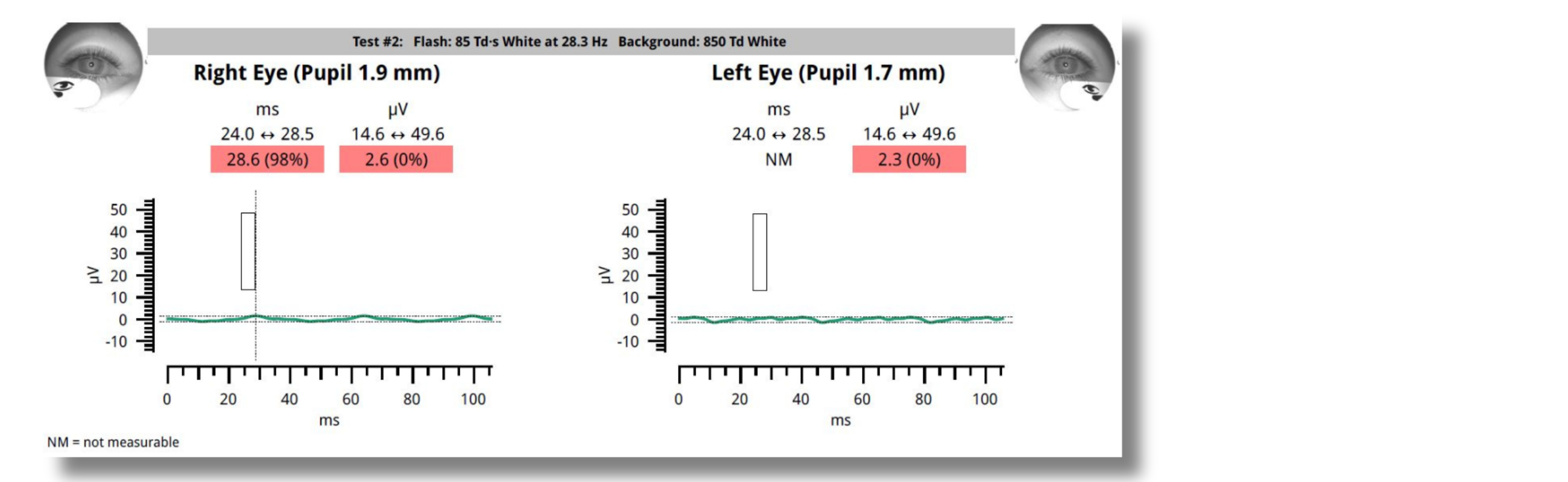
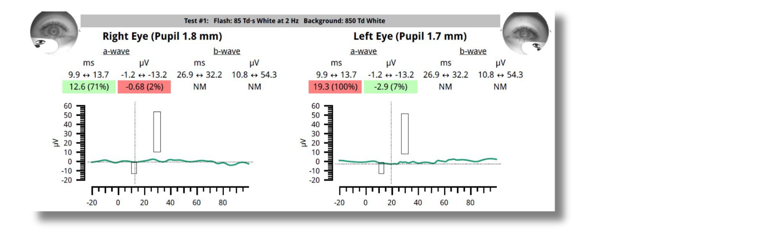
A photopic electroretinogram (ERG) was performed, which demonstrated abnormal cone function (Figure 3). Although not shown here, the scotopic ERG also revealed abnormal findings. These results are suggestive of underlying retinal degeneration.
Figure 3: ERG
How Did the ERG Impact Next Steps?
The patient was scheduled for genetic testing but was already informed of our preliminary diagnosis of atypical retinitis pigmentosa. She was also counseled regarding low vision support and available rehabilitation options.
Why We Use RETeval
ERG testing performed earlier could have prevented the cost and potential risks associated with refractive laser treatment, and it would have allowed for earlier education and management for her progressive vision loss.

Dr. Hussein Almuhtaseb
The View Hospital, Qatar
Dr. Hussein Almuhtaseb is a distinguished Consultant Ophthalmologist specializing in Cataract and Vitreoretinal Surgery, whose career is marked by clinical excellence and a forward-looking commitment to technological advancement in medicine.rnrnFollowing his residency at Sagrat Cor University Hospital in Barcelona Spain, Dr. Almuhtaseb further honed his expertise by completing four prestigious fellowships in Medical and Surgical Retina at leading institutions, including the University Hospital Southampton and the Manchester Royal Eye Infirmary. He subsequently served as a Consultant Ophthalmologist in Cataract and Vitreoretinal Surgery at North Manchester. In 2022, he established and led the Ophthalmology Department at The View Hospital in Doha, Qatar, a facility affiliated with Cedars-Sinai, solidifying his reputation as a leader in the field.rnrnIn addition to his surgical practice, Dr. Almuhtaseb holds a special interest in the application of Artificial Intelligence (AI) in Retina. He has actively pursued this passion, completing a certificate in AI in Healthcare: From Strategies to Implementation from Harvard University, positioning him at the intersection of advanced surgical technique and cutting-edge medical technology. His focus is on leveraging AI to enhance diagnostic accuracy, predict disease progression, and optimize treatment pathways for complex retinal conditions.