Estudo de Caso
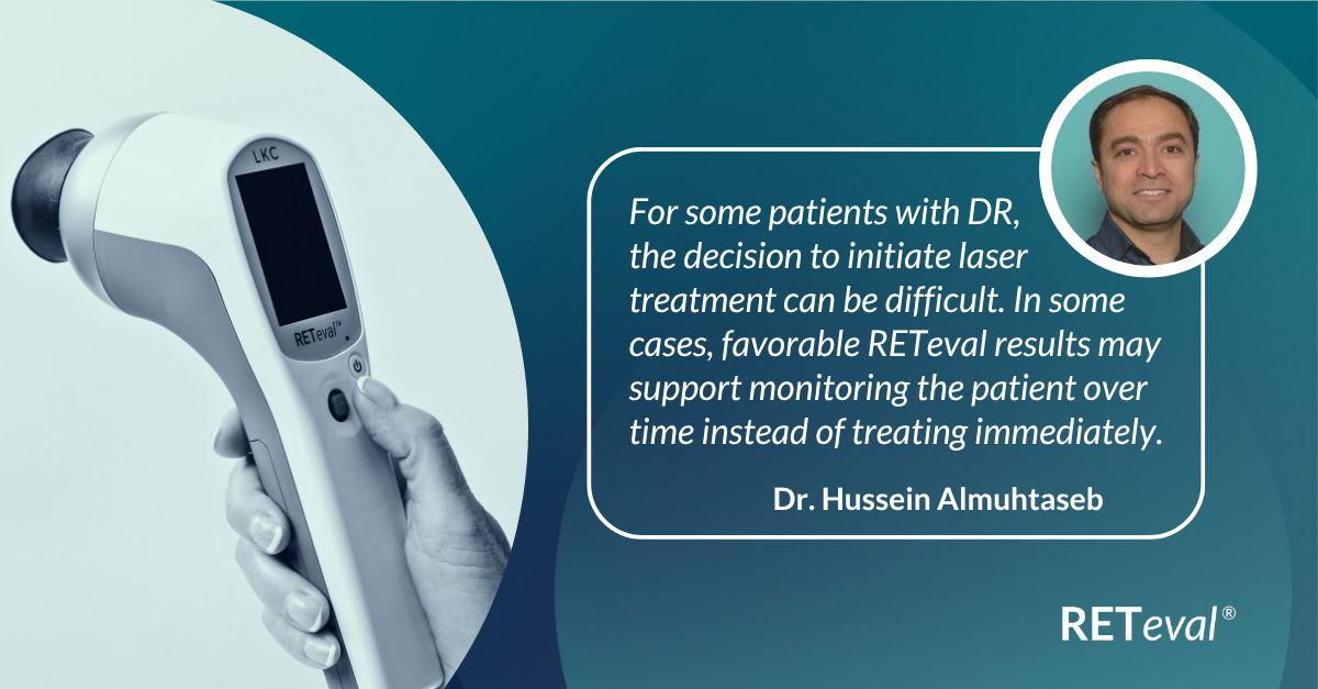
ERG Supports Treatment Decision in Diabetic Retinopathy
by Dr. Hussein Almuhtaseb, The View Hospital, Qatar

Desagio
Validating Delay of Treatment

Diagnóstico
Retinopatia Diabética

Testing Protocol
DR Assessment
Patient History
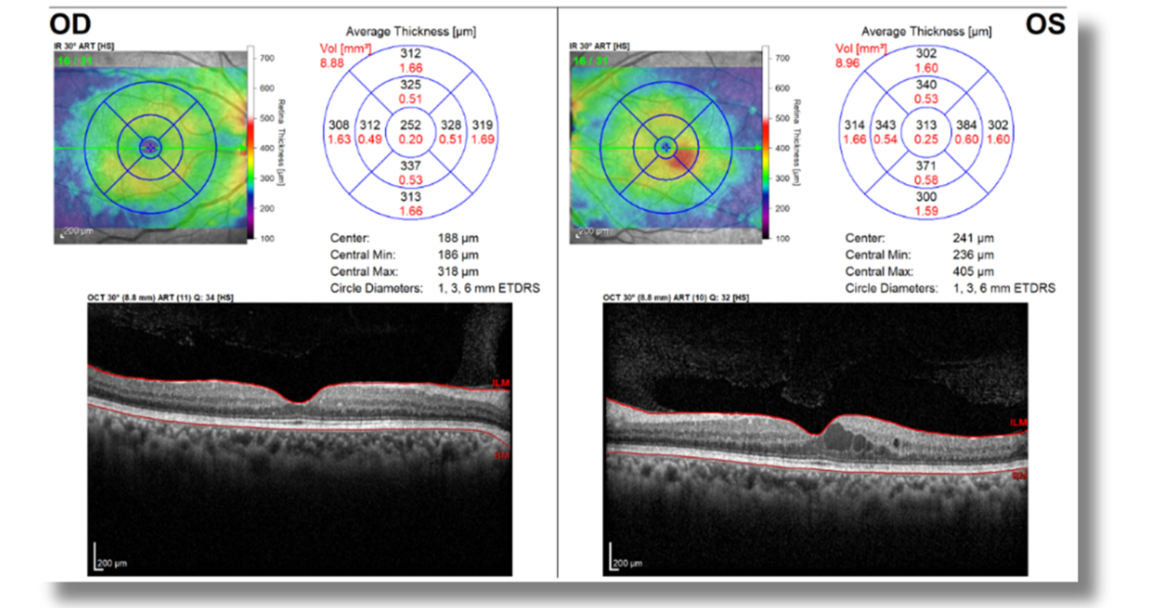
A 46-year-old patient with diabetic retinopathy and bilateral blurry vision presented to our clinic. Visual assessment revealed best corrected visual acuity of 0.8 in the left eye and 1.0 in the right eye. The left eye had a history of prior panretinal photocoagulation (PRP), and OCT findings (Figure 1) indicated mild diabetic macular edema (DMO). Given the history of PRP in the left eye, additional PRP was considered in response to signs of mild ischemia. However, the patient questioned the recommendation after experiencing loss of peripheral vision after PRP in the left eye.
Figure 1: OCT
Why Was an ERG Test Performed?
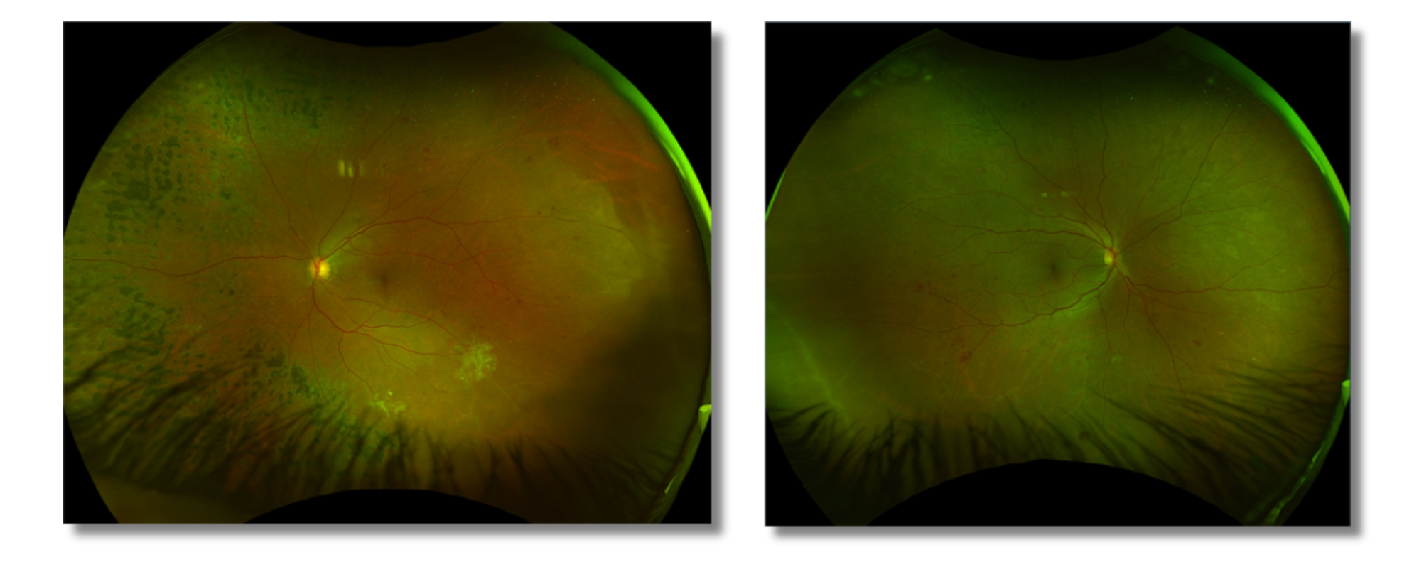
Fundus imaging findings (Figure 2) suggested the need for peripheral laser treatment; however, the signs were mild, and the patient expressed concerns about potential peripheral vision loss associated with PRP. As a result, a RETeval® ERG was requested to evaluate retinal function and help determine the risk of disease progression.
Figure 2: Fundus Images
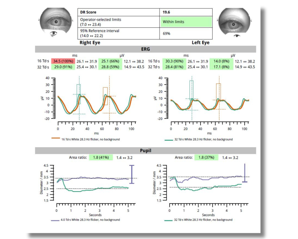
What Were the ERG Findings?
Although some changes were seen in the peripheral retina, the RETeval DR Score was 19.6. Based on the recent study by Davis et al., a score of 19.6 is linked to a 0% risk of progression at both 6 and 12 months, indicating very mild changes and low concern for disease progression. The ERG flicker results demonstrated amplitudes within normal limits, and the implicit times for the brighter flicker stimulus were also within the normal range. The reduced ERG amplitude in the left eye may be attributable to prior PRP treatment and a solid DR Score may indicate stable disease.
Figure 3: ERG Results
How Did the ERG Impact Next Steps?
The ERG results were reassuring, indicating that photocoagulation treatment was not necessary at that time. We opted for close monitoring to detect any signs of deterioration or unexpected developments. More than a year later, the eye remains stable without the need for PRP.
Why We Use RETeval
Using objective, functional testing to complement structural imaging ensures that we have the whole story and can make accurate disease management decisions. In this case, adding ERG with the RETeval gave us data to support not rushing into PRP for the right eye.

Dr. Hussein Almuhtaseb
The View Hospital, Qatar