
ERG, Explained
Why is structural testing alone not sufficient for retinal disease management?
Two patients can look nearly identical on an OCT, but they may progress very differently. Why? Structural imaging only shows the anatomy of the retinal tissue and is subjective to several factors, including patient compliance. Retinal dysfunction can also precede structural damage.1 That’s why functional assessments, such as electroretinography (ERG), are an important component of the ophthalmic diagnostic suite—to help providers identify disease and categorize their high-risk patients before complications develop.2
O que é eletrorretinografia (ERG)?
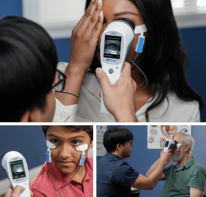
You may have heard of an ECG (electrocardiogram), which is a test that measures the electrical activity of the heart. An ERG, or electroretinogram, works similarly for the retina.
How does an ERG test work?
During an electroretinogram (ERG test), patients’ eyes are stimulated with light. When this stimulus hits the back of the eye, the resulting electrical activity is measured by skin or corneal electrodes. The way the retina responds can provide vital, objective information about a patient’s retinal function and health.
Are there different types of ERG tests?
There are 3 main types of ERG tests, each with different applications and benefits. Retinal function testing with these various types of ERG gives clinicians, researchers, and veterinary ophthalmologists a way to reliably monitor retinal changes or disease progression over time.

What is full-field ERG (ffERG)?
Full-field ERG uses a summed electrical response from a range of different cells and regions in the retina, revealing the full picture of a patient’s retinal health. The portable, handheld RETeval device is capable of performing this type of ERG test.

What is pattern ERG (PERG)?
During a pattern ERG test, an alternating checkerboard pattern is used as the stimulus to read electrical responses from the ganglion cells that compose the inner retina. This type of ERG is most commonly used to test for disorders affecting the ganglion cells and some neurodegenerative disorders.

What is multifocal ERG (mfERG)?
A multifocal (mfERG) test is performed with a similar protocol to a PERG, but the light stimulus displays as an array of hexagons that flicker from black to white in a pseudo-random sequence. mfERG is best used to measure central retinal dysfunction.
What can functional testing reveal that imaging may miss?
Functional testing may reveal changes sooner than they appear on imaging, such as optical coherence tomography (OCT) or fundus photography.1 Electroretinography objectively evaluates retinal function, while structural imaging shows the anatomy of the retinal tissue.
| ERG – functional retinal assessment: | Fundus, OCT, etc. – structural retinal assessments: |
| • Measures how well the retina responds to a light stimulus • Tracks electrical impulses that transmit visual information to the brain |
• Provides magnified images of the retina • Allows for assessment of retinal anatomy below the surface |
Why function and structure sometimes disagree
While both functional and structural assessments have their benefits, they can sometimes provide very different information. Retinal function changes can appear well before structural changes3, making a patient appear stable when they may already be at risk of progressing to advanced disease.
Combining both structural and functional assessments can help tell the full story of retinal health, making it easier for eyecare providers to catch critical abnormalities more quickly,2 helping them maximize patient vision retention.
O que é um eletrorretinograma?
The electroretinogram (ERG) is an examination that evaluates the function of the eye’s retinal cells. Patients’ eyes are stimulated with light and the resulting electrical activity from their retinal cells is measured by skin or corneal electrodes. Dependent on the type, intensity and colour of the light stimulus information on different areas and types of cells can be obtained.
Detectar o estresse funcional
Antecede o dano estrutural.
Qual a diferença entre o ERG e os testes estruturais de imagen?
A eletrorretinografia avalia objetivamente as anormalidades funcionais da retina, enquanto a imagem estrutural mostra a anatomia do tecido retiniano. Embora as avaliações funcionais e estruturais tenham seus benefícios, as mudanças funcionais geralmente aparecem bem antes das mudanças estruturais. Detectar anormalidades da retina rapidamente é fundamental para minimizar danos e maximizar a retenção da visão, por isso avaliações funcionais como o ERG são ferramentas importantes no conjunto de diagnósticos oftalmológicos.
Handheld ERG como uma ferramenta de diagnóstico prática e eficaz
Recent studies comparing ERG to structural imaging techniques show that ERG is an effective diagnostic tool. In studies comparing ERG and structural imaging’s abilities to evaluate sight-threatening diabetic retinopathy, the handheld and portable RETeval device’s ERG results outperformed the traditionally-used imaging technique in predicting which patients would later need medical intervention.[13],[14] Em uma avaliação da capacidade do RETeval’s ability to evaluate diabetic retinopathy, the advantages of RETeval incluíram detecção mais precoce da disfunção retiniana, menores custos de investimento e menos conhecimento de leitura necessário do que a técnica de imagem tradicionalmente usada.[15] Uma avaliação da capacidade do RETeval’s ability to evaluate central retinal vein occlusion found that RETeval ERGs são menos invasivos, mais fáceis de executar e fornecem previsões mais confiáveis do que a técnica de imagem tradicionalmente usada.[16]
Vantagens dos testes ERG
A facilidade e a sensibilidade dos ERGs realizados com o RETeval portátil são altamente preferidas em relação às técnicas de imagem em determinadas situações. Pacientes que apresentam opacidades da média, pouca cooperação ou pupilas pequenas são mais capazes de serem avaliados para anormalidades retinianas com o RETeval[14].
Teste ERG não invasivo
Em um estudo independente comparando RETeval ERGs com fotografias retinianas digitais convencionais, o RETeval foi escolhido pelos pacientes como o método preferido em relação à fotografia.[14] Embora muitas crianças precisem ser sedadas para se submeterem ao ERG convencional, crianças de até seis meses de idade têm sido capazes de permanecer calmas durante os ERGs realizados com RETeval.[18] A natureza não invasiva do ERG e a facilidade do RETeval portátil e não midriático tornam o dispositivo ERG uma ferramenta de triagem valiosa para muitas patologias oftálmicas.
RETeval, testes ERG simplificados
Aplicações da eletrorretinografia
A eletrorretinografia tem uma variedade impressionante de aplicações em humanos e animais, tanto para uso clínico quanto para pesquisa. Nossa tecnologia oferece soluções de ERG confiáveis, repetíveis e eficazes em todas essas categorias.


More about the RETeval ERG/VEP device
Popular Topics: Make a Difference in Diabetic Retinopathy Care | Glaucoma Evaluation with RETeval PhNR Test | RETeval Device Reference Data | RETeval in Optometry
Case Studies: When Standard Tests Aren’t Enough: Using ERG to Support Diagnosis in Case with High Myopia | DR Score Helps Overcome Blind Faith in Popular Weight Loss Drug |A Family Affair: ERG-Guided Care for a Married Couple | DR Score Prompts Critical Referral | A Tale of Two Patients: How ERG Uncovered Hidden Differences |ERG Supports Treatment Decision in Diabetic Retinopathy | ERG Demonstrates Stable Function Despite Severe Structural Damage | ERG Supports Treatment Decision in Diabetic Retinopathy | Photopic Negative Response as a Reliable Method for Glaucoma Follow-up in Children | A Tale of Two Patients | Vision Complaints Reflected on ERG | Predictive Value of Combining Diagnostic Technologies| ERG Provides Clarity When Fields and OCT Are Inconclusive| ERG Raises Red Flag, Changing Management Trajectory | ERG Provides Confidence to Monitor or Treat | ERG to Determine Ischemic Status | ERG to Replace FA for CRVO Treatment Decision | Using ERG to Monitor Glaucoma | Routine ERG Use Supports Complex Patient Management | ERG Alters Follow-up Schedule and Education for Patient with Diabetes | Using ERG for Management of Birdshot Chorioretinopathy | Using ERG to Monitor Glaucoma | Comprehensive Pediatric Assessment Using ERG in Challenging Cases | ERG Above and Beyond Retinal Imaging | ERG’s Role in Diabetic Retinopathy Progression Monitoring | Avaliação de Risco para CRVO baseado em ERG | ERG Supports Diagnostic Accuracy in a Pediatric Patient
Ebooks: How Modern ERG Became Essential for Diabetic Management | Core Cases from the Clinical Compendium | Modern Fundamentals of Diabetic Retinopathy Management in Optometry | Elevating Patient Care with ERG
Articles: Electroretinography Added to AAO’s Diabetic Retinopathy Preferred Practice Pattern Guidelines | How Comfortable is the RETeval for Patients? |The Use of RETeval ERG/VEP in Pediatric Ophthalmology | How RETeval ERG Has Enhanced My Practice | The Use of RETeval ERG/VEP in Pediatric Ophthalmology | The Ultimate Guide to Diabetic Retinopathy in Primary Eyecare | What Type of Functional Testing Do You Prefer for Patients with Diabetes? | Major Milestone: RETeval Referenced in over 200 Publications | Collaboration to Elevate the Standard of Care for DR | Is ERG Needed if You Have Access to a Good Structural Imaging Device? | A Straightforward Approach to Managing and Supporting Patients with Diabetes | Simplify Grading and Risk Assessment in Diabetic Retinopathy | Simplify Daily Decision-Making with Modern ERG | Objective Functional Testing Needs in Diabetes and Glaucoma | Why Modern ERG is Re-Defining Diabetes Management | Diabetic Retinopathy Management Protocols for Optometry
Videos: RETeval: Helping You Assess Progression Risk | RETeval: Eliminating Confusion in Clinic | RETeval: More Information, Better Decisions | RETeval: Eliminating Confusion in Clinic | RETeval: Function to Rely On | RETeval Handheld ERG: Features & Benefits | RETeval: Enhancing Collaborative Care | Handheld ERG for Primary Eyecare | Advice for Optometric Colleagues about Handheld ERG | Making a Difference in Diabetic Retinopathy Care | Changing the Way We Think About Electrodiagnostics | ERG Testing Made Simple | VEP Testing Made Simple | ERG Waveform | Introduction to Visual Electrophysiology | A Superior DR Progression Risk Assessment with the RETeval dispositivo | Improve Glaucoma Management with the RETeval Handheld ERG Device | Reshaping the Retinal Diagnostic Landscape | New Solutions for Infants with ROP | Using the RETeval in Myopia Research
Webinars: ABCs of ERG | ERG in the Exam Lane | Ready for RETeval | ERG in Action | Blueprint for Functional Assessments | Predicting Vision Loss in a Busy Retina Practice | Objective, Functional Testing for Glaucoma? | Best Management Practices for Diabetic Retinopathy | How the RETeval Device Became a Daily Instrument in my Diagnostic Toolkit