Estudo de Caso
ERG Supported Cataract Treatment Decision in Dementia Patient, Improving Vision and Emotional Well Being
by Dr. Hussein Almuhtaseb, The View Hospital, Qatar


Desagio
Support Disease Management Decisions

Diagnóstico
Cataract

Testing Protocol
ISCEV Flash/Flicker
Patient History
A 72-year-old male patient with dementia and a history of diabetes presented with significant visual impairment. The family reported challenges in daily care, including increased episodes of aggression and withdrawal—symptoms that had noticeably intensified in parallel with his declining vision. The patient had no light perception in the right eye and markedly reduced vision in the left. Best corrected visual acuity in the left eye was 0.2, improving slightly to 0.3 with pinhole testing. Examination revealed a dense, brunescent cataract in the left eye, with severely limited fundus visibility, as illustrated in the accompanying images (Figure 1). The family and patient were primarily concerned about whether cataract extraction could meaningfully improve his residual vision.
Figure 1: OCT & Fundus

Why Was an ERG Test Performed?
Fundus imaging was inconclusive due to poor media clarity. Previous consultants had discouraged cataract surgery, citing a high-risk profile and uncertain benefit in a cognitively impaired and behaviorally challenging patient. However, the profound burden placed on both the patient and his caregivers due to the vision loss warranted a deeper investigation. To better understand the functional capacity of the retina and to support an informed decision, an electroretinogram (ERG) was performed.
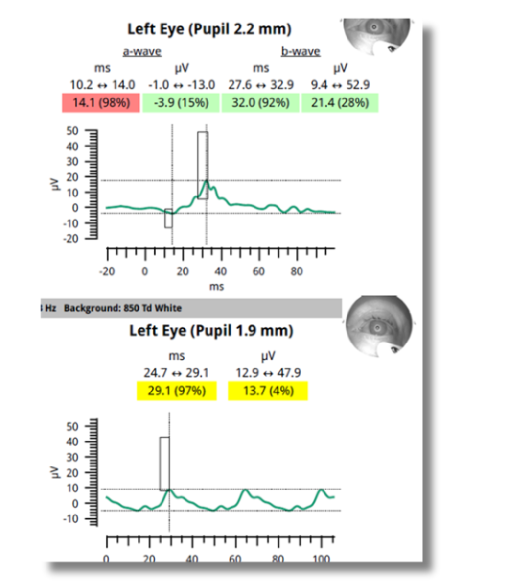
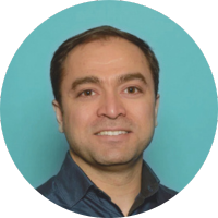
What Were the ERG Findings?
The ERG revealed amplitudes and implicit times within borderline to normal ranges (Figure 2), which represents an encouraging result. This suggested that despite the media opacity, the retinal function remained largely intact. Based on these findings, we communicated to the patient and family that there is a reasonable likelihood of visual improvement following cataract surgery.
Figure 2: ERG
How Did the ERG Impact Next Steps?
Cataract surgery was performed on the left eye, resulting in an improved visual acuity of 0.7 postoperatively. The patient is being closely monitored to manage potential ocular effects related to his diabetes. At one of the postoperative visits, the family reported a striking and unexpected transformation: the patient’s mood had brightened, and his level of engagement had noticeably increased. With better vision, he appeared more responsive, more curious, and more connected to his surroundings, which is a huge relief for the family. The impact of improved vision extended beyond measurable acuity.
Why We Use RETeval
Had the ERG not been performed, a definitive recommendation for surgery may not have been possible. This uncertainty could have led to deferral of treatment, leaving the patient in a state of visual and emotional stagnation. In this case, the ERG not only informed a successful surgical decision, but ultimately contributed to remarkable improvements in both visual function and emotional well-being—reminding us of the profound interconnection between vision, cognition, and quality of life.
Dr. Hussein Almuhtaseb
The View Hospital, Qatar
Dr. Hussein Almuhtaseb is a distinguished Consultant Ophthalmologist specializing in Cataract and Vitreoretinal Surgery, whose career is marked by clinical excellence and a forward-looking commitment to technological advancement in medicine.
Following his residency at Sagrat Cor University Hospital in Barcelona Spain, Dr. Almuhtaseb further honed his expertise by completing four prestigious fellowships in Medical and Surgical Retina at leading institutions, including the University Hospital Southampton and the Manchester Royal Eye Infirmary. He subsequently served as a Consultant Ophthalmologist in Cataract and Vitreoretinal Surgery at North Manchester. In 2022, he established and led the Ophthalmology Department at The View Hospital in Doha, Qatar, a facility affiliated with Cedars-Sinai, solidifying his reputation as a leader in the field.
In addition to his surgical practice, Dr. Almuhtaseb holds a special interest in the application of Artificial Intelligence (AI) in Retina. He has actively pursued this passion, completing a certificate in AI in Healthcare: From Strategies to Implementation from Harvard University, positioning him at the intersection of advanced surgical technique and cutting-edge medical technology. His focus is on leveraging AI to enhance diagnostic accuracy, predict disease progression, and optimize treatment pathways for complex retinal conditions.