Case study
ERG-based Risk Assessment in CRVO
by Patrícia de Freitas Dotto, MD, PhD,
Dr. Jeser Amarante Faria Children’s Hospital, Joinville - Brazil
An 87-year-old female patient sought medical attention due to gradual vision loss. She presented with a two-month history of blurred vision OD, a dense cataract and 20/200 best corrected visual acuity (BCVA). Following examination and imaging, she was diagnosed with central retinal vein occlusion (CRVO) with diffuse edema.

Challenge
Dense Cataract

Diagnosis
CRVO

Type of protocol
Flicker test
Why was the ERG test performed?
Image quality was borderline low through the cataract, yet we needed to evaluate disease severity more closely in order to determine whether the condition was ischemic or non-ischemic and to assess the patient’s risk of developing rubeosis iridis. A quickly to conduct flicker ERG test can indicate if CRVO is ischemic or non-ischemic and help assess risks and complications associated with CRVO. [59]
Learn more about the use of the RETeval ERG/VEP device:
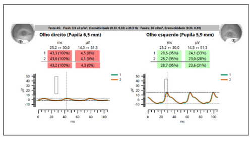
What were the ERG findings?
The flicker ERG results revealed significantly delayed implicit time and low amplitude in the affected eye, as well as a large inter-eye difference. In sum, these results indicated that the patient has ischemic CRVO and is at high risk to progress to rubeosis iridis.
How did ERG influence the patient’s care?
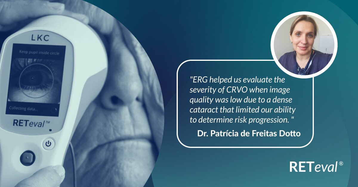
The patient underwent cataract surgery with vitrectomy as well as panretinal photocoagulation (PRP) treatment to avoid progression to rubeosis. The patient now has a stable retinal appearance and no change in best corrected visual acuity (BCVA) of the affected eye. ERG helped us evaluate the severity of CRVO when image quality was low due to a dense cataract.
Conclusion
The dense cataract negatively influenced image quality, impacting our ability to provide an accurate assessment. An ERG test can be successfully completed even if media opacities are present. In this case, ERG played a crucial role in identifying the ischemic nature of CRVO and predicting the patient’s high risk of developing rubeosis iridis.


Patrícia de Freitas Dotto
Hospital Infantil Jeser Amarante Faria – Joinville, Santa Catarina- Brazil
Dr. Patrícia de Freitas Dotto has dedicated a large part of her life to ophthalmology and clinical research along with participating in a visual electrophysiology scholarship program at Escola Paulista de Medicina – Federal University of São Paulo. Dra. Patricia’s expertise in electrophysiology guided the subsequent assessment and management of this complex case.