Case study
Using ERG to Monitor Glaucoma
by Dr. Edith Rom, Augenarztpraxis Dr. Domscheit & Dr. Neißkenwirth, Germany
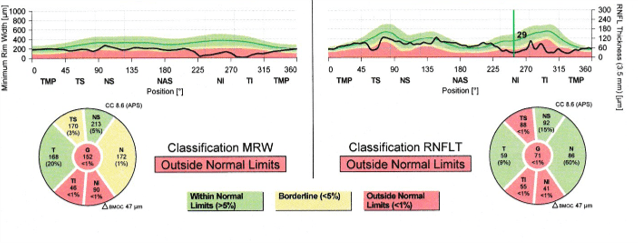
A 62-year-old female presented with high myopia, low-tension glaucoma with recurrent disc margin hemorrhages, progressive thinning of the optic nerve rims, and visual field defects. She received multiple treatments over a 3-month period. These included topical IOP-lowering medications, acupuncture, and laser trabeculoplasty.

| ONH (OD) | ONH (OS) |
|---|---|
 |
 |
Why was the ERG test performed?
Establishing treatment success in patients with progressive glaucoma is very challenging. In this case, the optic-disc cupping was continuously worsening, which made it difficult to determine treatment effect. The ERG provided a means to confirm whether or not the treatment plan was having a positive impact.
What were the ERG findings?
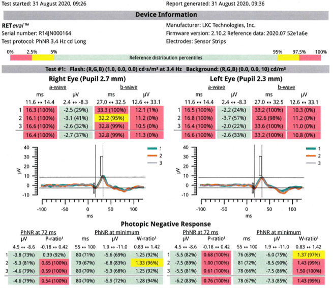
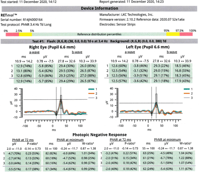
Just before we initiated treatment, the PhNR test parameters, as measured by the RETeval device, were outside the normal limits. After three months, all values improved and fell within the recommended reference ranges. Correspondingly, the patient also had slight contrast sensitivity improvements.
| ERG (PhNR Protocol) before treatment | ERG (PhNR Protocol) after treatment |
|---|---|
 |
 |
How did ERG influence the patient’s care?
The ERG findings provided peace of mind that we are on the right treatment path, but need to continue to closely monitor both structural and functional parameters in the event her condition worsens and requires additional intervention, such as surgery.
Conclusion
This case demonstrates how ERG provides useful information about changes to ganglion cell function that help us more easily monitor treatment effects. Having this additional sensitive parameter helps us decide if and when additional measures are needed or if the current approach is adequate in the short-term.


