ASSESS PROGRESSION RISK
A Single Score for Diabetic Retinopathy
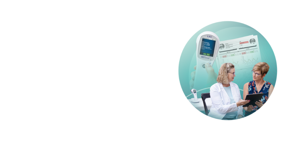
Give your patients the advantage of timely, actionable information with the only handheld ERG that delivers an objective diabetic retinopathy exam: the RETeval®.
How do you know which of your patients will progress? How do you know when?
Traditional imaging may not reveal functional damage until it’s too late, especially in non-proliferative stages. That means missed opportunities for timely diabetic retinopathy treatment and better patient outcomes.
The RETeval Solution
- Science-backed DR Score – Stratify progression risk and determine which patients need more of your time and attention
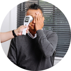
- Non-invasive – No dilation, no corneal contact
- Efficient – Test in minutes, train staff quickly, integrate into any workflow
- Versatile – Ideal for primary and specialty care
Backed by science
Landmark Research
The RETeval handheld ERG’s objective DR Score has been validated by more than 20 peer-reviewed publications since 2016. Explore the full research library here.
2025
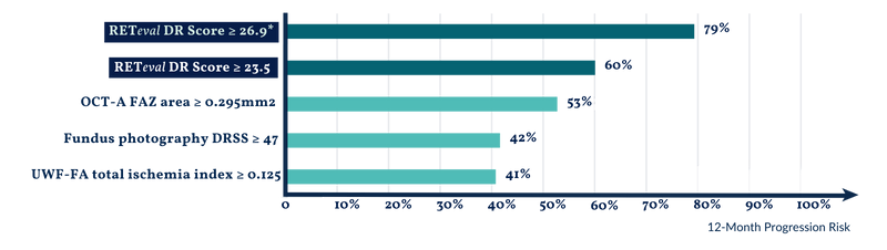
RETeval DR Score ≥ 26.9 predicts 79% chance for progression from moderate to severe NPDR to vision-threatening complications in less than 1 year.
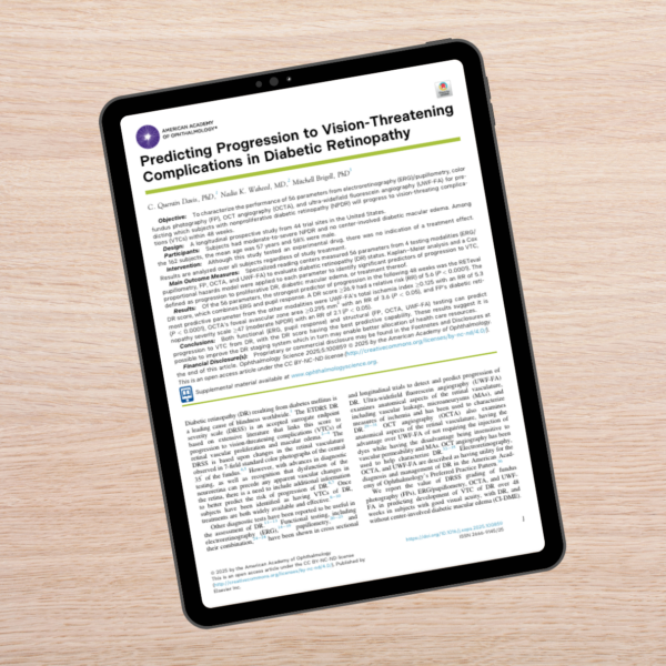
Davis Q, Waheed N, Brigell M: Predicting Progression to Vision-Threatening Complications in Diabetic Retinopathy. Ophthalmology Science. June 2025.
2020
Each 1-point change in the DR Score increases the probability of progression from severe NPDR to ocular intervention by 28% over 3 years.
Brigell M, Chiang B, Maa A, Davis Q: Enhancing Risk Assessment in Patients with Diabetic Retinopathy by Combining Measures of Retinal Function and Structure. Translational Vision Science & Technology. August 2020.
2016
Confirms RETeval’s efficacy in detecting vision-threatening diabetic retinopathy.
Maa A et al: A novel device for accurate and efficient testing for vision-threatening diabetic retinopathy. Journal of Diabetes and Its Complications. December 2015.
Why the DR Score Matters
Latest research confirms a RETeval DR Score ≥ 26.9 outperforms 55 diagnostic parameters on 4 testing modalities, including OCT-A, ultra-widefield fluorescein angiography, and fundus photography in predicting progression to vision-threatening complications within 1 year.
- Allows targeted referrals and/or resource prioritization, reducing overtreatment and missed progression
- Creates potential to improve DR staging systems and support value-based care models
- Supports evidence for inclusion of ERG in the American Academy of Ophthalmology’s updated Preferred Practice Pattern for detecting and managing DR
upcoming webinar
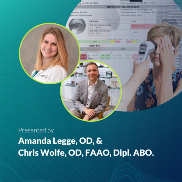
Data That Makes a Difference
Wednesday, July 22
8:00 PM Eastern (5:00 PM Pacific)
- Which of these patients should come back sooner?
- How do I motivate this patient to make important changes?
- Does my patient need to see a retina specialist?
Every day, eyecare providers find themselves asking questions just like these as they navigate conflicting clinical data and demanding schedules. Assessing patients’ risk for disease progression without the help of objective, functional data can feel like reading a map with missing pages.
If this sounds familiar, join us for Data that Makes a Difference, a peer-to-peer webinar led by practicing optometrists Amanda Legge, OD, and Chris Wolfe, OD, FAAO, Dipl. ABO.
Study confirms: RETeval DR Score outperforms structural testing for DR risk assessment
Diabetic retinopathy is the leading cause of blindless worldwide—and your patients are counting on you to help preserve their sight with all the tools available to help you assess their risk for disease progression.
A November 2025 study from Ophthalmology Science found that a RETeval DR Score of 26.9 or higher outperformed three forms of structural testing—OCT-A, ultra-widefield FA, and fundus photography—in identifying which patients with moderate to severe NPDR would progress to vision-threatening complications within 1 year.
RETeval: Making a Difference in Diabetic Retinopathy Care
designed for clinical reality
Why RETeval?

handheld, lightweight, portable
Easy to use anywhere in your practice

NO CORNEAL CONTACT
LKC patented Sensor Strips keep testing comfortable.

TEST WITHOUT DILATION
Real-time pupillography automatically adjusts for pupil size

seamless integration
Train techs in minutes thanks to on-demand clinical resources and ongoing support
ERG Named in Preferred Practice Pattern Guidelines for Diabetic Retinopathy
The American Academy of Ophthalmology’s inclusion of ERG demonstrates its valuable role in both diagnosing and managing diabetic retinopathy. This decision reflects the growing recognition that objective, functional testing, alongside structural imaging, is critical for a comprehensive DR assessment.
See why clinicians are calling this a pivotal moment in eye care history >
“The inclusion of ERG in the AAO’s Preferred Practice Pattern guidelines is a pivotal moment in eye care history. As diagnosticians and treating physicians, we need an objective, functional complement to structural imaging. ERG provides this, helping us detect early retinal dysfunction that may precede visible structural changes.”

see reteval in action
Case Studies & Clinical Insights
A Tale of Two Patients: How ERG Uncovered Hidden Differences
The Predictive Value of Combining Diagnostic Technologies
ERG’s Role in Diabetic Retinopathy Progression Monitoring
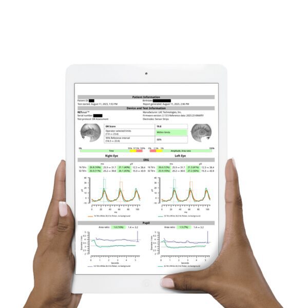
RETeval DR Assessment Reports
Easy-to-interpret, color-coded reports with:
- Age-adjusted reference data
- Science-backed DR Score
- Functional stress measurements
- Pupil response metrics
Ready for more?
Protect vision. Improve outcomes. Predict the future of diabetic eye disease.