Case study
ERG’s Role in Diabetic Retinopathy Progression Monitoring
by Dr. Pamela Weber, Island Retina, Shirley, USA
A 39-year-old patient presented with a 20-year history of type 1 diabetes mellitus. The initial examination revealed nonproliferative diabetic retinopathy (NPDR) with diabetic macular edema (DME). In response to these findings, anti-VEGF injections were administered to address the diabetic macular edema. The RETeval ERG/VEP was used at follow-up exams from 2017 through 2019 to aid in monitoring disease progression, ultimately alerting us to advancement to PDR and a need for more aggressive treatment.

Challenge
Early recognition of disease progression

Diagnosis
NPDR with DME

Type of protocol
DR Assessment
Why was the ERG test performed?
To monitor changes and possible disease progression in order to make informed decisions regarding the need for more aggressive treatment.
What were the ERG findings?
Despite no significant changes on OCT or fundus photography over the years, ERG revealed a deterioration, prompting fluorescein angiography examination, which in turn revealed neovascularization elsewhere (NVE) as well as progression from NPDR to PDR.
| 2017 | Several injections and focal laser in both eyes. The DR Score is relatively high (24.3 for 23.4 limit), with low amplitudes. | 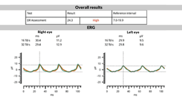 |
|---|---|---|
| 2018 | The DR Score remains unchanged, but amplitudes improve significantly. | 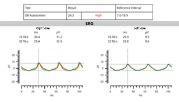 |
| 2019 | A remarkable decrease in amplitude in the left eye, even though the fundus and OCT image didn’t show significant change. | 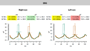 |
| ERG results indicate the need for close monitoring to watch for ischemia, invisible on fundus, but appearing on FA. Neovascularization elsewhere (NVE) was detected in the periphery and the eye was treated more aggressively. | 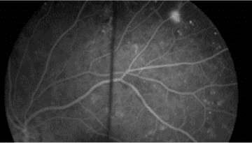 |
How did ERG influence the patient’s care?
The ERG offered an efficient method for monitoring a high-risk patient over several years. Concerning ERG findings demonstrated a need to schedule more frequent follow-up appointments and also prompted more extensive testing, including FA. This revealed NVE, and a progression from NPDR to PDR with ischemia. Accordingly, the patient received more aggressive treatment.
Conclusion
This case highlights the crucial role of ERG in monitoring progression in patients with diabetic retinopathy, particularly in cases resistant to treatments. The use of ERG with other diagnostic tools can facilitate a more complete understanding of the clinical picture, reducing delays in necessary follow-up exams and treatment.
Learn more about the use of the RETeval ERG/VEP device in measuring diabetic retinopathy progression risk:

Pamela Weber, MD
Island Retina, Shirley, NY, USA
Dr. Pamela Ann Weber is a retinal specialist with Vitreoretinal Consultants of New York, USA, and practices at the Island Retina office in Shirley. She specializes in retinal disease assessment, including diabetic retinopathy. Dr. Weber has a rich academic background and extensive experience. She received her B.Sc. from McGill University, Montreal, and her MD from Columbia College of Physicians and Surgeons, NYC. She completed her residency in ophthalmology at the New York Eye and Ear Infirmary, and a fellowship in retina at Harvard University, Boston. Dr. Weber utilizes the RETeval ERG/VEP device for monitoring retinal conditions in her practice.