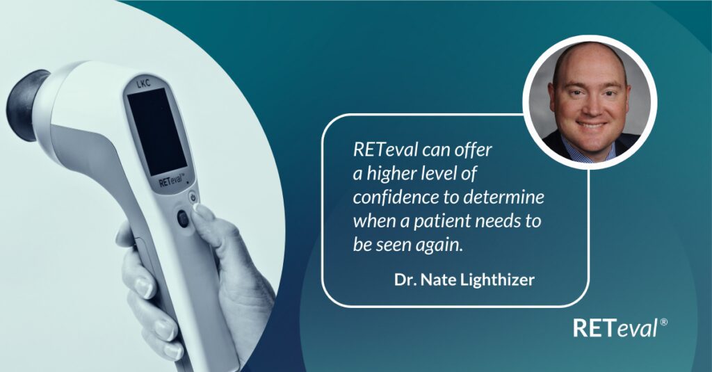
ERG to Determine Ischemic Status
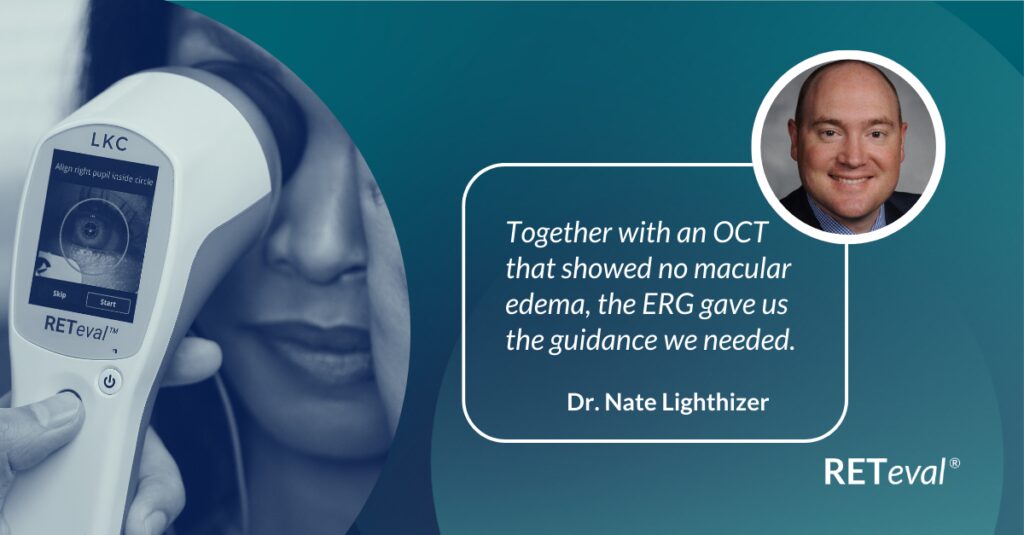
Explore how Dr. Nate Lighthizer treated a 50-year-old female patient who initially presented for punctal plugs. Although she wasn’t scheduled for a retinal evaluation, a resident noticed reduced vision in her left eye.
ERG to Replace FA for CRVO Treatment Decision
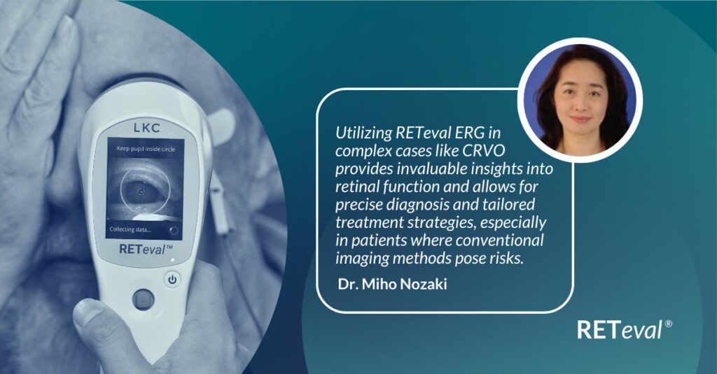
Dr. Miho Nozaki demonstrates the value of RETeval ERG for diagnosing CRVO in complex cases, where conventional testing poses risks.
Using ERG to Monitor Glaucoma
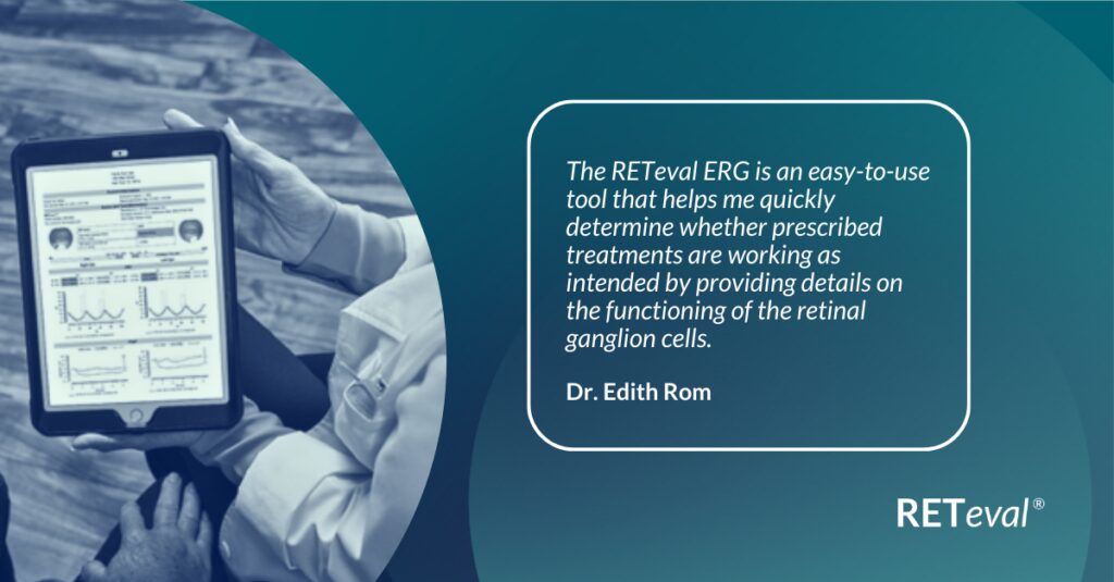
Explore the use of Electroretinography (ERG) in managing Glaucoma through a case study by Dr. Edith Rom, Germany.
ERG Alters Follow-up Schedule and Education for Patient with Diabetes
A 33-year-old Native American female presented because she lost her glasses a year prior. From a visual standpoint, she was doing well. She didn’t report any changes in her vision and was corrected to 20/20 in both eyes (-1.25 OD, -1.00 OS). Her last A1c was 10.0, which is certainly higher than we would like to see.
Using ERG for Management of Birdshot Chorioretinopathy
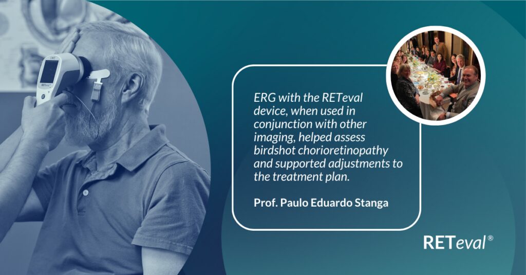
Explore the use of Electroretinography (ERG) in managing Birdshot Chorioretinopathy through a detailed case study by Prof. Paulo Eduardo Stanga and team at The Retina Clinic London. This study examines a 62-year-old patient’s journey from initial diagnosis to advanced treatment, including intravitreal therapy and immunosuppressive therapy.
Comprehensive Pediatric Assessment Using ERG in Challenging Cases

Learn how Dr. Mario Zanolli utilized ERG to assess a 4-year-old boy with nystagmus when standard imaging techniques proved challenging. This case study demonstrates the effectiveness of ERG in pediatric cases where traditional methods may not provide clear results.
Routine ERG Use Supports Complex Patient Management
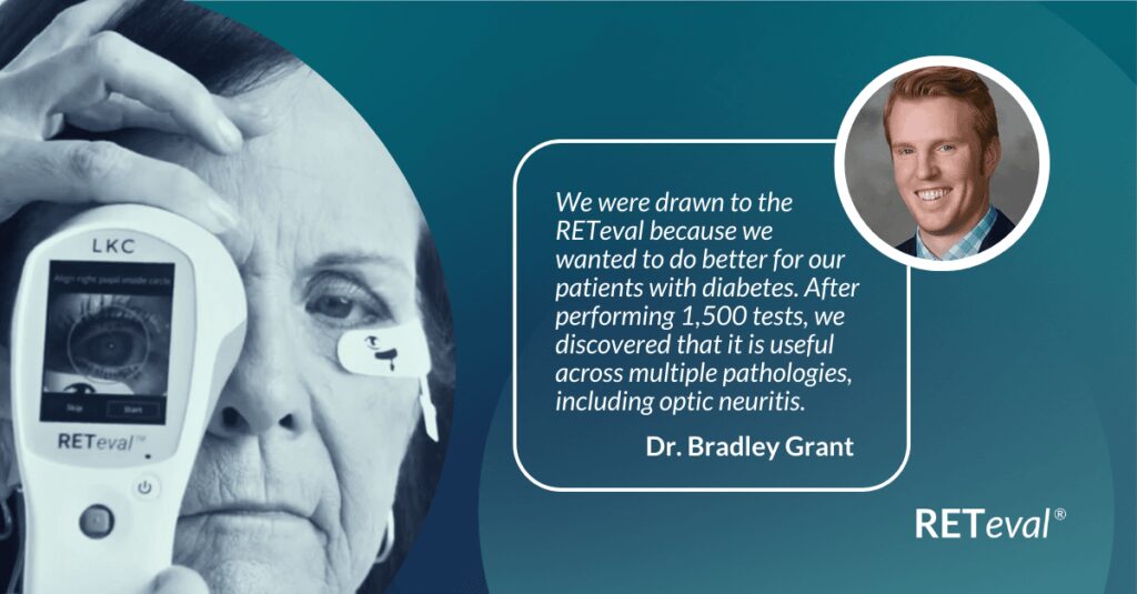
Subjective functional assessments don’t always yield useful information. Bradley Grant, an optometrist in Shelbyville, IL, utilized his RETeval handheld ERG to determine the appropriate follow-up schedule for a patient with multiple sclerosis, while leveraging the easy-to-interpret, objective functional data to educate the patient on the importance of resuming care with their neurologist.
ERG Above and Beyond Retinal Imaging
A 7-year-old child was referred to confirm a multiple sclerosis (MS) diagnosis following a brain scan. The patient complained of visual changes but identifying the cause was complex due to limited patient cooperation. We obtained color fundus and OCT images, both of which appeared to be normal. A flash visual evoked potential (VEP) test was conducted at another clinical site, but the outcome was unreadable.
ERG’s Role in Diabetic Retinopathy Progression Monitoring

Explore how Dr. Pamela Weber used the RETeval ERG/VEP to monitor the progression of diabetic retinopathy in a 39-year-old patient with type 1 diabetes. The case study shows how early detection and monitoring with advanced retinal imaging can lead to timely and more aggressive treatment, improving patient outcomes.
ERG-based Risk Assessment in CRVO
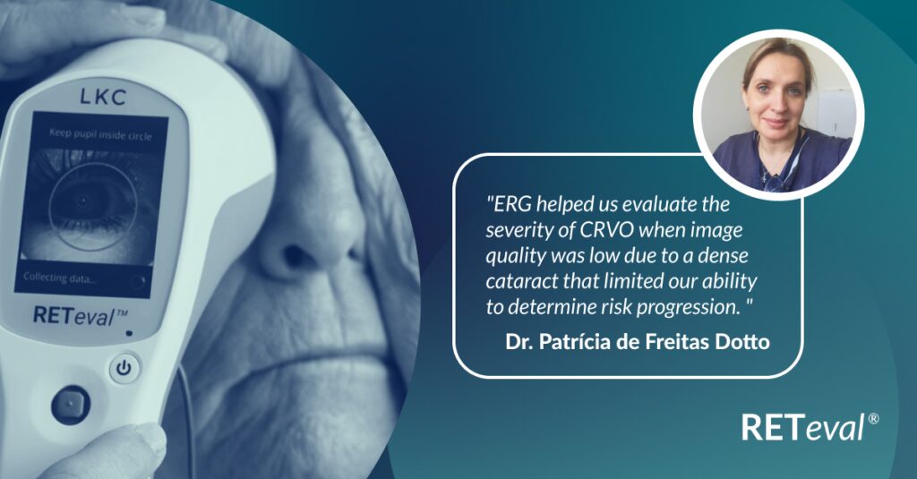
This case study involves an 87-year-old female patient sought medical attention due to gradual vision loss. She presented with a two-month history of blurred vision OD, a dense cataract and 20/200 best corrected visual acuity (BCVA).
ERG Supports Diagnostic Accuracy in a Pediatric Patient
This case study involving a 3-year-old boy with bilateral high myopia (-6.25D and -7.00D), normal structural results, and no extraocular features demonstrates how ERG addresses these challenges.