
Electroretinography and Visual Evoked Potential
Go Beyond Structure
Measure Function
Achieve superior patient outcomes and power your research with ERG/VEP testing for an objective, more complete understanding of retinal function.
Ophthalmic Veterinarian
Why Run ERG or VEP Tests?
ERG testing (electroretinography) and VEP testing (visual evoked potential) solutions play a vital role in obtaining diagnostic information in both humans and animals, aiding clinicians and researchers with comprehensive data on how the retina and visual pathways are functionally operating.

DIAGNOSE
Efficient and effective ERG / VEP testing provides an objective aid in detecting and confirming the presence of inherited or acquired eye disease.

MONITOR
Track disease progression and/or predict treatment needs and outcomes.

RESEARCH
Expand your understanding of retinal function and assess the factors impacting retinal processes and visual pathways with accurate and repeatable results.
Our ERG/VEP Devices
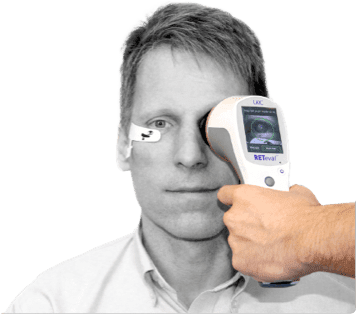
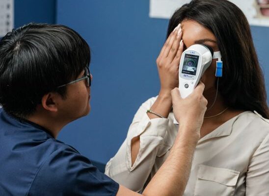
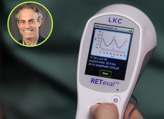
RETeval®
The only FDA-cleared, portable, battery-powered, non-mydriatic ERG/VEP Testing Device. ISCEV-compliant. Remarkably straightforward to use.
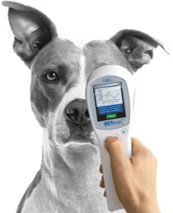
RETevet™
For both veterinary ophthalmologists and researchers, measure an animal’s retinal function with a view of the eye in real-time.
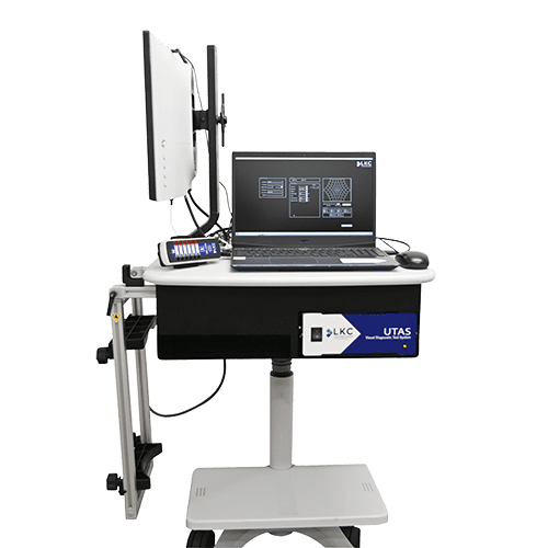
UTAS™ mf/PERG
Compact and comprehensive testing for multifocal and pattern ERG and VEP assessments. Intuitive interface and fully ISCEV-compliant.
Need supplies?
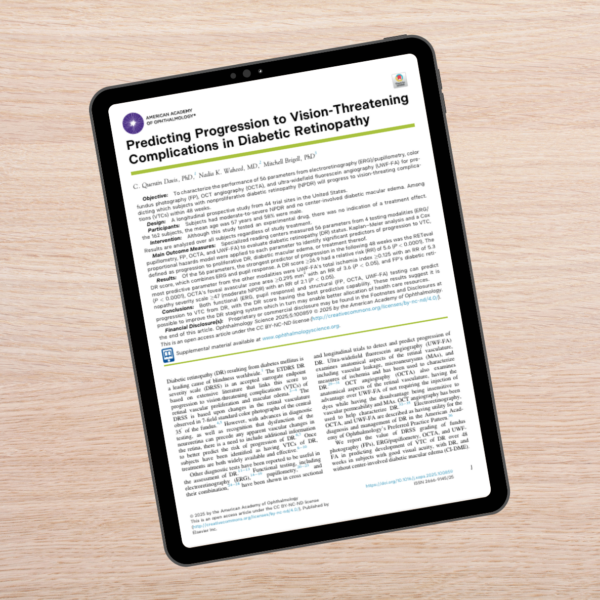
Study confirms: RETeval DR Score outperforms structural testing for DR risk assessment
Diabetic retinopathy is the leading cause of blindless worldwide—and your patients are counting on you to help preserve their sight with all the tools available to help you assess their risk for disease progression.
A November 2025 study from Ophthalmology Science found that a RETeval DR Score of 26.9 or higher outperformed three forms of structural testing—OCT-A, ultra-widefield FA, and fundus photography—in identifying which patients with moderate to severe NPDR would progress to vision-threatening complications within 1 year.
upcoming webinar
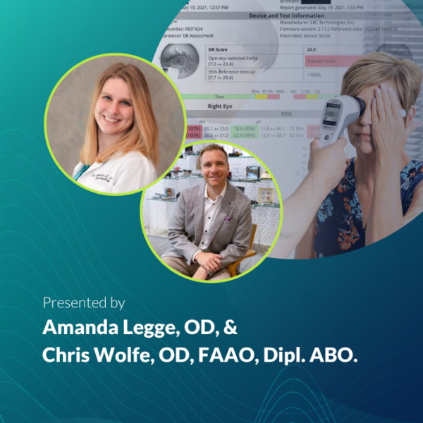
Data That Makes a Difference
Wednesday, July 22
8:00 PM Eastern (5:00 PM Pacific)
- Which of these patients should come back sooner?
- How do I motivate this patient to make important changes?
- Does my patient need to see a retina specialist?
Every day, eyecare providers find themselves asking questions just like these as they navigate conflicting clinical data and demanding schedules. Assessing patients’ risk for disease progression without the help of objective, functional data can feel like reading a map with missing pages.
If this sounds familiar, join us for Data that Makes a Difference, a peer-to-peer webinar led by practicing optometrists Amanda Legge, OD, and Chris Wolfe, OD, FAAO, Dipl. ABO.
“We have been very pleased with the RETeval. It has changed the way that we practice and has allowed us to significantly decrease the amount of EUAs with ERGs we perform. Furthermore, it has resulted in earlier diagnosis of patients with inherited retinal degenerations.”

ERG Named in Preferred Practice Pattern Guidelines for Diabetic Retinopathy
The American Academy of Ophthalmology’s inclusion of ERG demonstrates its valuable role in both diagnosing and managing diabetic retinopathy. This decision reflects the growing recognition that objective, functional testing, alongside structural imaging, is critical for a comprehensive DR assessment.
See why clinicians are calling this a pivotal moment in eye care history >
Webinars
These webinars offer tips and comprehensive information on why electrophysiology and LKC’s systems and devices are the perfect solution for you.
The Blueprint for Functional Assessments
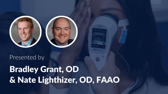
How can ERG predict vision loss in a busy retina practice?

New to Functional Testing? Here are the ABCs of ERG.
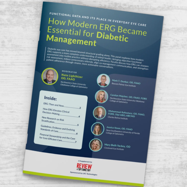
How Modern ERG Became Essential for Diabetic Management
First published in Review of Optometry, this cohort of nationally recognized optometrists and educators discuss how modern ERG has evolved from a specialized test to a practical tool that supports everyday management for patients with diabetes.
Two patients can look nearly identical on imaging, yet progress very differently. Modern ERG adds objective functional data to help clarify risk, reduce uncertainty, and guide more precise follow-up and referral decisions. The result is clearer clinical decision-making and more actionable patient conversations.
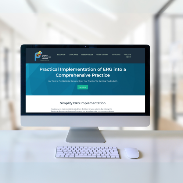
Practical Implementation of ERG into a Comprehensive Practice
Chris Wolfe & EyeCode Education
With this course by Chris Wolfe, OD, FAAO, Dip. ABO, available from Practice Performance Partners, you’ll know exactly when and how to use ERG to improve patient care and protect your practice. We’re giving away coupon codes so you can take the course for FREE!
“RETeval really has made me a better diagnostician. It has allowed me to get early information about a patient’s eye health before disease becomes clinically visible.”


About LKC
LKC Technologies® is a 35-year veteran of visual electrophysiology (ERG). The RETeval ERG is the only handheld, FDA-cleared device for ERG testing on dilated and un-dilated patients. It was designed to streamline clinic operations and simplify testing for staff and patients alike. It is supported by more than 250 peer-reviewed publications and is used in optometry, ophthalmology, and research settings.
Latest Articles
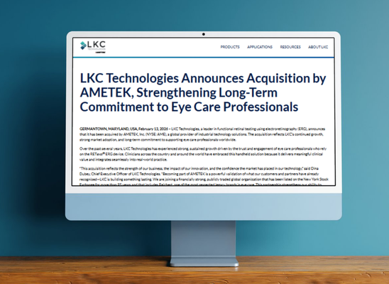
AMETEK Acquires LKC Technologies
Retinal Physician, February 2026
LKC Technologies has been acquired by AMETEK as part of a strategic move to expand their ophthalmic portfolio. The acquisition complements AMETEK’s existing eyecare diagnostics business, including Reichert.
Expanding ERG in Diabetic Eye Care
The Ophthalmologist, November 2025
Recent evidence shows the RETeval DR Score is the strongest predictor of progression to vision-threatening complications, enabling more proactive, personalized care.
Handheld RETeval device outshines other metrics for predicting DR complications
Healio, October 2025
Could a single number help predict vision-threatening diabetic retinopathy? New research from Ophthalmology Science finds the RETeval DR Score outperformed other metrics in predicting progression to vision-threatening complications.
Latest Research
Use of the RETeval Handheld Electroretinogram Device for Assessing the Risk of Diabetic Retinopathy in Patients With Type 1 Diabetes: A Case Report
November 19, 2025
Sirek, Sebastian, Barbara Trepka-Sirek, Sebastian Seget, Aleksandra Górska, Przemysława Jarosz-Chobot, und Dorota Pojda-Wilczek. „Use of the RETevalTM Handheld Electroretinogram Device for Assessing the Risk of Diabetic Retinopathy in Patients With Type 1 Diabetes: A Case Report“. Cureus, Online-Vorab-Publikation, 19. November 2025.
Assessment of Retinal Function Using Full-Field Electroretinography in Patients Undergoing Vitrectomy for a Macular Hole
November 20, 2025
Górska, Aleksandra, Sebastian Sirek, und Dorota Pojda-Wilczek. „Assessment of Retinal Function Using Full-Field Electroretinography in Patients Undergoing Vitrectomy for a Macular Hole“. Cureus, Online-Vorab-Publikation, 20. November 2025.
More about the RETeval ERG/VEP device
Popular Topics: Make a Difference in Diabetic Retinopathy Care | Glaucoma Evaluation with RETeval PhNR Test | RETeval Device Reference Data | RETeval in Optometry
Case Studies: ERG Demonstrates Stable Function Despite Severe Structural Damage | ERG Supports Treatment Decision in Diabetic Retinopathy | Photopic Negative Response as a Reliable Method for Glaucoma Follow-up in Children | A Tale of Two Patients | Vision Complaints Reflected on ERG | Predictive Value of Combining Diagnostic Technologies| ERG Provides Clarity When Fields and OCT Are Inconclusive| ERG Raises Red Flag, Changing Management Trajectory | ERG Provides Confidence to Monitor or Treat | ERG to Determine Ischemic Status | ERG to Replace FA for CRVO Treatment Decision | Using ERG to Monitor Glaucoma | Routine ERG Use Supports Complex Patient Management | ERG Alters Follow-up Schedule and Education for Patient with Diabetes | Using ERG for Management of Birdshot Chorioretinopathy | Using ERG to Monitor Glaucoma | Comprehensive Pediatric Assessment Using ERG in Challenging Cases | ERG Above and Beyond Retinal Imaging | ERG’s Role in Diabetic Retinopathy Progression Monitoring | ERG-based Risk Assessment in CRVO | ERG Supports Diagnostic Accuracy in a Pediatric Patient
Ebooks: Core Cases from the Clinical Compendium | Modern Fundamentals of Diabetic Retinopathy Management in Optometry | Elevating Patient Care with ERG
Articles: Electroretinography Added to AAO’s Diabetic Retinopathy Preferred Practice Pattern Guidelines | How Comfortable is the RETeval for Patients? |The Use of RETeval ERG/VEP in Pediatric Ophthalmology | How RETeval ERG Has Enhanced My Practice | The Use of RETeval ERG/VEP in Pediatric Ophthalmology | The Ultimate Guide to Diabetic Retinopathy in Primary Eyecare | What Type of Functional Testing Do You Prefer for Patients with Diabetes? | Major Milestone: RETeval Referenced in over 200 Publications | Collaboration to Elevate the Standard of Care for DR | Is ERG Needed if You Have Access to a Good Structural Imaging Device? | A Straightforward Approach to Managing and Supporting Patients with Diabetes | Simplify Grading and Risk Assessment in Diabetic Retinopathy | Simplify Daily Decision-Making with Modern ERG | Objective Functional Testing Needs in Diabetes and Glaucoma | Why Modern ERG is Re-Defining Diabetes Management | Diabetic Retinopathy Management Protocols for Optometry
Videos: RETeval: More Information, Better Decisions | RETeval: Eliminating Confusion in Clinic | RETeval: Function to Rely On | RETeval Handheld ERG: Features & Benefits | RETeval: Enhancing Collaborative Care | Handheld ERG for Primary Eyecare | Advice for Optometric Colleagues about Handheld ERG | Making a Difference in Diabetic Retinopathy Care | Changing the Way We Think About Electrodiagnostics | ERG Testing Made Simple | VEP Testing Made Simple | ERG Waveform | Introduction to Visual Electrophysiology | A Superior DR Progression Risk Assessment with the RETeval Device | Improve Glaucoma Management with the RETeval Handheld ERG Device | Reshaping the Retinal Diagnostic Landscape | New Solutions for Infants with ROP | Using the RETeval in Myopia Research
Webinars: ABCs of ERG | Ready for RETeval | ERG in Action | Blueprint for Functional Assessments | Predicting Vision Loss in a Busy Retina Practice | Objective, Functional Testing for Glaucoma? | Best Management Practices for Diabetic Retinopathy | How the RETeval Device Became a Daily Instrument in my Diagnostic Toolkit