RETEVAL DEVICE FOR ELECTRORETINOGRAPHY
Revolutionary ERG/VEP
Anywhere for Anyone
Enhance your diagnostic capabilities or fuel your research with the RETeval® device — a powerful aid in the diagnosis and management of retina and optic nerve diseases such as diabetic retinopathy, glaucoma, and inherited retinal dystrophies.
Why RETeval?
For Clinicians
For Researchers

ERG Named in Preferred Practice Pattern Guidelines for Diabetic Retinopathy
The American Academy of Ophthalmology’s inclusion of ERG demonstrates its valuable role in both diagnosing and managing diabetic retinopathy. This decision reflects the growing recognition that objective, functional testing, alongside structural imaging, is critical for a comprehensive DR assessment.
See why clinicians are calling this a pivotal moment in eye care history >
VISUAL ELECTROPHYSIOLOGY TESTING
Detect Functional Stress
Anticipate Structural Damage
ERG and VEP tests provide objective information on the function of the visual system. It gives reliable guidance for medical professionals to manage functional changes that may impact a patient’s vision, typically in advance of structural changes.
Explore ERG Explore VEP
“We have been very pleased with the RETeval. It has changed the way that we practice and has allowed us to significantly decrease the amount of EUAs with ERGs we perform. Furthermore, it has resulted in earlier diagnosis of patients with inherited retinal degenerations.”

The RETeval device helps doctors to:
1. DETECT
Detect various retina and optic nerve diseases earlier
2. PREDICT
Predict the progression of the disease
3. FOLLOW
Then follow up on the course of disease
4. MONITOR
Monitor treatment success

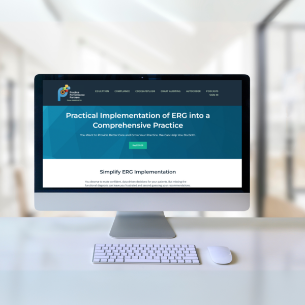
Practical Implementation of ERG into a Comprehensive Practice
Chris Wolfe & EyeCode Education
With this course by Chris Wolfe, OD, FAAO, Dip. ABO, available from Practice Performance Partners, you’ll know exactly when and how to use ERG to improve patient care and protect your practice. We’re giving away coupon codes so you can take the course for FREE!
Explore Applications
ERG and VEP play a vital role in obtaining diagnostic information in both humans and animals, aiding clinicians and researchers with comprehensive data on how the retina and visual pathways are functioning.

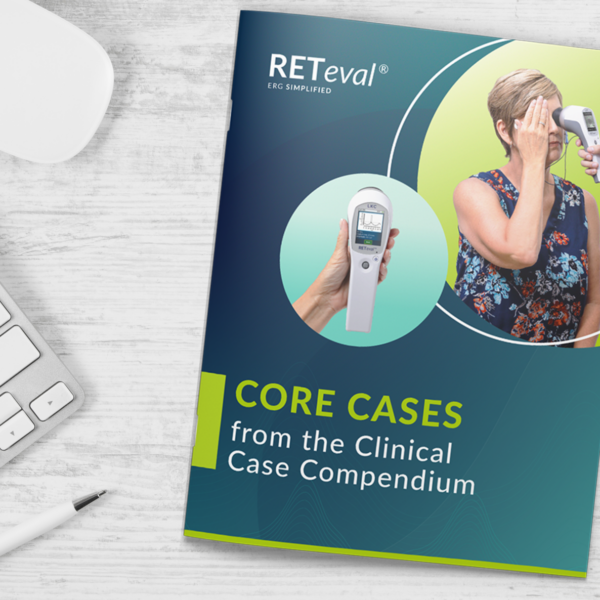
RETeval: Core Cases from the Clinical Compendium
With the incorporation of a single simple test, these eyecare providers are elevating their patient care through easier management decisions and better patient education.
With each of the 4 cases presented in this eBook, learn:
-
- Why was an ERG performed?
- What were the ERG findings?
- How did the ERG impact next steps?

SIMPLE TO INTERPRET
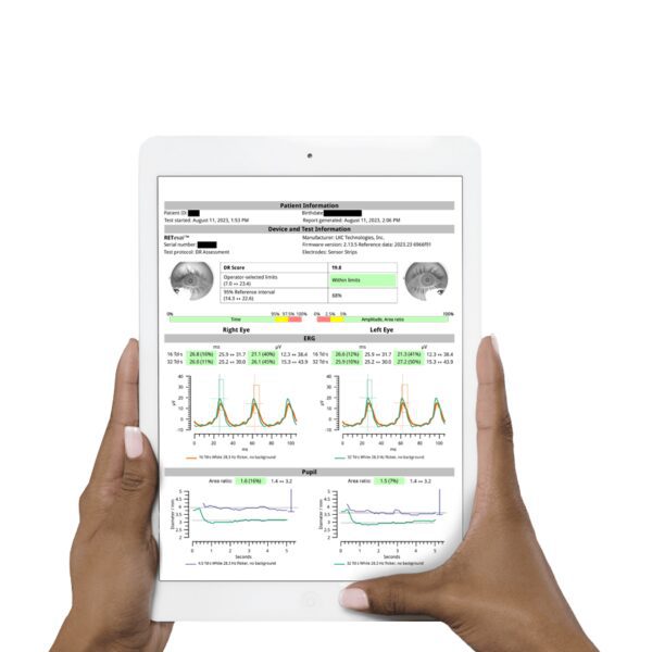
RETeval Reports
Color-coded results compared to an age-adjusted reference data set make interpretation straightforward and efficient in a busy practice. Results are easily exported into any EMR/EHR system.
SAMPLE REPORTS
Diabetic Retinopathy Assessment
PhNR
ERG & VEP Coding and Reimbursement Guides
In the United States, there are more than 560 ICD-10-CM codes that may be associated with the CPT codes used to report electroretinography and visual-evoked potential. The following guides, developed by The Pinnacle Health Group, provide some of the more common diagnosis codes that may be used for protocols associated with the RETeval handheld device.
Download ERG guide Download VEP guide
The above guides focus on Physician Office Coding and Reimbursement. For Hospital Outpatient, contact us.
“RETeval really has made me a better diagnostician. It has allowed me to get early information about a patient’s eye health before disease becomes clinically visible.”


How It Works
-
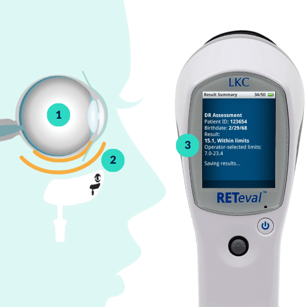
The RETeval device starts flashing light into the patient’s eye.
-
The retina responds to the flashes by generating small electrical signals that travel through the facial structure to the Sensor Strip.
-
The Sensor Strip detects the electrical signals and compares the results to the age-adjusted reference database.
RETeval, ERG Testing Made Simple
RETeval Resources
RETeval Brochure (US)
RETeval Brochure (International)
Data Barcode App
ERG Coding & Reimbursement Guide (US)
CA Prop 65 Warning
Order Sensor Strips
Enhancing Risk Assessment in Patients with Diabetic Retinopathy by Combining Measures of Retinal Function and Structure
August, 2020
Brigell M, Chiang B, Maa A, Davis Q. Translational Vision Science & Technology. 2020; 9(9):40.
Screening for diabetic retinopathy in diabetic patients with a mydriasis-free, full-field flicker electroretinogram recording device
November 12, 2019
Zeng Y, Cao D, Yu H, et al. British Journal of Ophthalmology 2019;103:1747-1752.
Constant luminance (cd·s/m2) versus constant retinal illuminance (Td·s) stimulation in flicker ERGs
February 3, 2017
Davis Q, Kraszewska O, Manning C. Documenta Ophthalmologica. 2017: 134, 75–87.
More about the RETeval ERG/VEP device
Popular Topics: Make a Difference in Diabetic Retinopathy Care | Glaucoma Evaluation with RETeval PhNR Test | RETeval Device Reference Data | RETeval in Optometry
Case Studies: ERG Demonstrates Stable Function Despite Severe Structural Damage | ERG Supports Treatment Decision in Diabetic Retinopathy | Photopic Negative Response as a Reliable Method for Glaucoma Follow-up in Children | A Tale of Two Patients | Vision Complaints Reflected on ERG | Predictive Value of Combining Diagnostic Technologies| ERG Provides Clarity When Fields and OCT Are Inconclusive| ERG Raises Red Flag, Changing Management Trajectory | ERG Provides Confidence to Monitor or Treat | ERG to Determine Ischemic Status | ERG to Replace FA for CRVO Treatment Decision | Using ERG to Monitor Glaucoma | Routine ERG Use Supports Complex Patient Management | ERG Alters Follow-up Schedule and Education for Patient with Diabetes | Using ERG for Management of Birdshot Chorioretinopathy | Using ERG to Monitor Glaucoma | Comprehensive Pediatric Assessment Using ERG in Challenging Cases | ERG Above and Beyond Retinal Imaging | ERG’s Role in Diabetic Retinopathy Progression Monitoring | ERG-based Risk Assessment in CRVO | ERG Supports Diagnostic Accuracy in a Pediatric Patient
Ebooks: Core Cases from the Clinical Compendium | Modern Fundamentals of Diabetic Retinopathy Management in Optometry | Elevating Patient Care with ERG
Articles: Electroretinography Added to AAO’s Diabetic Retinopathy Preferred Practice Pattern Guidelines | How Comfortable is the RETeval for Patients? |The Use of RETeval ERG/VEP in Pediatric Ophthalmology | How RETeval ERG Has Enhanced My Practice | The Use of RETeval ERG/VEP in Pediatric Ophthalmology | The Ultimate Guide to Diabetic Retinopathy in Primary Eyecare | What Type of Functional Testing Do You Prefer for Patients with Diabetes? | Major Milestone: RETeval Referenced in over 200 Publications | Collaboration to Elevate the Standard of Care for DR | Is ERG Needed if You Have Access to a Good Structural Imaging Device? | A Straightforward Approach to Managing and Supporting Patients with Diabetes | Simplify Grading and Risk Assessment in Diabetic Retinopathy | Simplify Daily Decision-Making with Modern ERG | Objective Functional Testing Needs in Diabetes and Glaucoma | Why Modern ERG is Re-Defining Diabetes Management | Diabetic Retinopathy Management Protocols for Optometry
Videos: RETeval: More Information, Better Decisions | RETeval: Eliminating Confusion in Clinic | RETeval: Function to Rely On | RETeval Handheld ERG: Features & Benefits | RETeval: Enhancing Collaborative Care | Handheld ERG for Primary Eyecare | Advice for Optometric Colleagues about Handheld ERG | Making a Difference in Diabetic Retinopathy Care | Changing the Way We Think About Electrodiagnostics | ERG Testing Made Simple | VEP Testing Made Simple | ERG Waveform | Introduction to Visual Electrophysiology | A Superior DR Progression Risk Assessment with the RETeval Device | Improve Glaucoma Management with the RETeval Handheld ERG Device | Reshaping the Retinal Diagnostic Landscape | New Solutions for Infants with ROP | Using the RETeval in Myopia Research
Webinars: ABCs of ERG | Ready for RETeval | ERG in Action | Blueprint for Functional Assessments | Predicting Vision Loss in a Busy Retina Practice | Objective, Functional Testing for Glaucoma? | Best Management Practices for Diabetic Retinopathy | How the RETeval Device Became a Daily Instrument in my Diagnostic Toolkit


